The Physical Therapy Homepage
Gait Analysis of Walking
By Dickie Gipson
Mechanical Kinesiology
November 20, 1996
Introduction
There are thousands of sports and activities that occur which would be impossible without the ability of walking. Walking is vital for ambulation and mobility of the human body. Development of walking, known as gait development, begins in pre-adolescence and continues to form and change throughout a person life. A person gait can become abnormal due to numerous factors related to malfunctions in the body. The development of an abnormal gait is caused by one or more of these malfunctions, and the body compensates for the malfunction by deviating the gait from the standard gait of a healthy body. A normal gait has been devised by doctors and is used to compare to a patient’s gait to aid in diagnosis of any abnormalities. This method of diagnosis is called gait analysis. To understand the uses and effectiveness of gait analysis, one must first understand the biomechanical intricacies involved in walking. This paper introduces and explains these intricacies by looking at the kinematics, by providing information from research conducted by numerous doctors, physical therapists, and other experts, and by summarizing all muscles that are used in walking. This paper also establishes a general knowledge of walking, gait analysis, and methods of physical therapy used to return people to a normal gait.
Kinematic Description
"Kinematics is the description of motion, including patterns and speed of movement sequencing by the body segments that often translates to degree of coordination an individual displays." (3, 3). The analysis of a motion begins with a description of the starting or initial position. The initial position of walking is with the legs even with one another and shoulder width apart, with the trunk up straight, and the arms down to the sides of the body. The action of walking can be divided into three basic phases. Phase One, known as the Swing Phase, involves lifting and extending the first leg forward, the dorsiflexing the ankle, and then extension of the ankle back
down to the floor. ( 7, 409-413) In this phase, the initial leg is only extended out about a shoulder width in front of the body in order not to over-extend the body’s center of gravity. If the leg was over-extended, the force of the extended leg would travel back toward the center of gravity and act as a retarding force in continuing the next phase of walking. This extension of the leg forward is known as a person’s stride. Also in this phase, when the first leg is lifted and extended, all weight is placed on the opposite leg and the opposite arm is swung forward slightly for three reasons. The first reason of swinging the arm forward is to act as a counter balance for the extension of the front leg. The second reason of swinging the arm is an indirect action of the rotation of the hips and pelvic girdle. The third reason is to use the force generated by the pulling back of the arm to the body to supply the force needed to extend the second leg.
The second phase of walking occurs in each leg and involves the flexing of the knee of the other leg and plantar flexion of the ankle. The weight of the body is transferred to the front leg in the second phase, which is also known as the stance phase. (5, 188) This transition is performed to enable continual movement of the hind or follow leg and position the body for the third phase.
The third phase involves bringing the hind leg forward in front of the body, extension of the knee back to the ground, and dorsiflexion of the ankle. The arms exchange position with the front arm pulled back to the body and the opposite arm is slightly extended forward. These three phases are repeated over and over in smooth fluent movement. Sequential motion and continual execution of these phases are needed to transfer the momentum that accumulates with each step. If the motion is not continual, then inertia must be overcame with each step, and a choppy gait will result. This concept involves the principles explained in Newton’s Law of Inertia. Newton’s 2nd Law states that an object at rest will remain at rest; An object in motion will remain in motion and travel in a straight line with uniform speed unless acted upon by an outside force. (3, 361)
Balance is vital in walking, and proper transfer of the body’s weight to each leg while the other leg is being moved forward is very important. Although brief descriptions of the body’s weight transfer is provided with each phase, a closer analysis can be deduced from the diagram in figure 1 that illustrates the application of the body’s weight during each phase of the walking or gait cycle. (5, 188)
 Figure 1: Body Weight Support in Relation to Balance
From the figure above, it can be seen that in the gait cycle there are two occasions where the body is supported by both feet, labeled ‘Double Support’ and colored yellow. There are also two instances when the weight is singularly supported or supported by only one foot. This is illustrated in green and each leg supports the weight while the opposite leg is in its swing phase. During these instances, the body shifts to that side of support in order to maintain balance and prevent falling. However, this shift is small and unnoticeable in a normal gait cycle, and the more fluent and faster, the less noticeable the shift become.
Another aspect to consider when analyzing the kinematics of a motion is the angular displacement of the body segments and the cardinal plane in which the motion is directed in. Angular displacement is the rotational motion that occurs in a body segment around an axis which is perpendicular to the motion. (3, 347) There are three cardinal planes in which the body moves within. The transverse plane divides the body into an upper and lower halves. (7, 10) The frontal plane divides the body into front and back halves. (7, 10) The sagittal plane divides the body into right and left halves. (7, 10) The sagittal plane is the cardinal plane in which the motions of walking are directed about. However, general angular displacement for the entire lower extremities is difficult to calculate due to the differences in the three joints which make up the lower extremities and origins for all angular motion. To obtain a better perspective of actual motions, analysis of each joint is more advantageous. Analysis of angular displacement in the
sagittal plane for the hip joint is illustrated by the graph in figure 2. (2, 214)
 Figure 2: Angular Displacement of the Hip in the Sagittal Plane
As can be seen in figure 2, the hip rests at approximately 80 in anatomical position. When the
hip is in flexion, the angle decreases to below 80 , and hip extension, the angle rises above 80 . Labeled A and C on the figure are the two occasions of flexion in the hip. These two flexions correspond to the two swing phases in the walking sequence. (5, 190) Labeled B on the graph is the point of extension which occurs during the stance phase. This correlation continues through
out each gait cycle.
 Angular Displacement of the Knee in the Sagittal Plane
Next, the angular displacement of the knee joint is analyzed and demonstrated in Figure 3. (2,214) As stated on the figure, the knee rests in anatomical position at 0 , with flexion being higher than 0 and extension being 0 or lower. Labels A, B, C, and D on the graph highlight the major motions performed by the knee joint. Label A represents a flexion of approximately 20 , which occurs in the stance phase of the gait cycle. (5, 190) The flexion occurs as a result of transferring the body’s weight to that limb. The knee flexes in order to absorb and support the gravitational force which acts upon the mass of the body. Label B marks a point of extension in the knee joint. This also occurs within the stance phase as the follow leg is extended forward in front the lead leg. Label C represents a 60 flexion of the knee joint. (5, 190) This flexion occurs during the swing phase as the knee is bent to bring the leg forward for the next step. Label D shows a return to extension in the swing phase and describes motion of the knee joint being straightened to perform the heel strike of the next step. (5, 190)
Finally, the angular displacement of the ankle joint in the sagittal plane is graphed and illustrated in figure 4. (2, 214)
 Figure 4: Angular Displacement of the Ankle in the Sagittal Plane
The motions of the ankles can be explain in four sections labeled A, B, C, and D on the graph above. Label A represents a dorsiflexion of the ankle as the heel is extended downward in order to make first contact on the floor and absorb the forces transferred to the foot. This motion is apart of the lead foot’s swing phase. Label B illustrates the plantar flexion involved in returning the ankle to a state in which the foot is flat upon the floor. This action is also within the swing phase but organizes and sets the lower extremities to enable the stance phase to occur. Label C demonstrates another period of dorsiflexion that occurs in the stance phase as the follow leg is brought forward. This causes the body to be positioned in front of the initial lead leg, and the angle between the tibia and the foot is diminished. Thus, a form of dorsiflexion results in the motion. Finally, in label C the second occurrence of plantar flexion occurs in the second swing phase. As the lead limb is brought forward, the ankle is extended down to prevent catching the heel on the floor.
All of these motions coincide to create a fluent and smooth pattern that is characteristic of a normal gait cycle. When the gait cycle is correctly utilized and repeated in a sequential manner, a good walking pattern or overall gait results. However, the normal gait is a very intricate and complex system. Any malfunction or debilitating trait of the human body can cause the gait to become abnormal.
Study of Literature
There are numerous doctors, physical therapists, and other professionals who have experimented and researched every aspect of the human gait and walking. This section summarizes sources that provide information such as the mechanical principles which are factors in the human gait, various malfunctions in the body that lead to a pathological gait, and a detailed study of gait analysis and the various factors to consider. Each of these topic areas has been broken down into subsections to provide better understanding and organization. Of some sources, too much material was available to express all contents so one major focal point from that material was emphasized and discussed in the appropriate section of interest.
Mechanical Principles
There are many mechanical principles involved in the human gait cycle. These principles are listed and their relativity to walking explained in the book, "Kinesiology" by Katherine F. Wells, Ph.D. These are the principles involved with gait. The first principle states, "Translatory movement of a lever is achieved by the repeated alternation of two rotatory movement, the lever turning first about one end, then about the other end." (7, 418) The gait cycle utilizes this principle with the rotation of the femur bone first at the hip joint and then at the knee joint.
The second principle says, "A body at rest will remain at rest unless acted upon by a force" (7, 418) This principle is utilized throughout the gait cycle. With the legs swing forward and being shifted to the back as the next step is taken, the legs act as pendulums attached to the rest of the body. If movement is not sequential, the momentive force from the previous step is lost and inertia must be overcame with every step.
The next principle explains that, "The speed of the gait is directly related to the magnitude of the pushing force and the direction of its application." (7, 418) This means the more force generated by the muscles of the lower extremities, the faster the gait cycle will be carried out and repeated. This also applies to increasing the speed of walking. The faster the gait cycle is carried out, the faster the pace of the person’s walk.
Another principle related to walking states, "Walking has been described as alternating loss and recovery of balance." (7, 418) This involves the swing phases of the gait cycle. As the leg is brought forward, the center of gravity is shifted forward and balance is lost. This is used to aid in propulsion of the limb forward, and once the leg has made contact with the surface, balance is recovered.
The next principle introduces that, "...propulsion of the body is effected by the diagonal pressure
of the foot against the supporting surface..." (7, 419) This principle takes into account Newton’s 3rd Law stating that with every force there is an equal but opposite reaction-force. Therefore, the supporting surface applies a reaction force equal but opposite to the force applied by the foot. Therefore, propulsion forward results from each push-off that occurs in the beginning of the swing phase.
The final principle involved with walking and the gait cycle is, "Stability of the body is directly related to the size of the base of support." (7, 419) The distance between the medial borders of the feet plays a major role in maintaining balance. Too small a distance between the feet causes a decrease in the base of support, and the stability is decreased as well. If the feet are spread far apart, the base of support is increased, and the body will be more stable. However, too wide a base of support while walking will hinder proper functions and produce a weaving, unsteady gait rather than a straight, fluent one. "The optimum position of the feet seems to be one in which the inner borders fall approximately along a straight line." (7, 419)
Reasons of Pathological Gait
There are many reasons for a person’s gait to be abnormal. The following gait abnormalities and the pathologies that cause them are listed and explained in the textbook called, "The Soft Tissues, Trauma and Sports Injuries" written by G. McLatchie and C. Lennox. In many forms of abnormal gait, the main cause is weakened muscles. The body compensates for this weakness, and the abnormal gait forms. The following abnormalities are results of weakened muscles or muscles that have atrophied.
Lateral trunk deviation involves the leaning of the trunk over the malfunctioning limb during the
stance phase to minimize the contraction of the gluteus medius muscle due to weakness or pain. Anterior and posterior trunk deviation also can cause the trunk to lean to compensate for weakened or injured muscles. Anterior trunk deviation involves weak knee extensors such as the Vasti group and Rectus Femoris. Posterior trunk deviation results from weakened hip extensors. (5, 191)
Functional leg length discrepancy occurs with the leg being at the wrong length during different stages of swing and stance phases. For example, the leg is not flexed in order to bring it forward or the leg is not extended to receive the body’s weight. This problem can lead to four different abnormal gait patterns. The first involves circumduction in which the swing out laterally to clear the ground. The second, vaulting, involves standing on the tiptoes to lengthen the stance leg. The third abnormality is called hip-hiking in which the pelvis is elevated and therefore elevates the swing leg to clear the ground. The final result of functional leg length discrepancy is steppage. Steppage involves the swing leg being shortened in the hip and knee by way of increased flexion. (5, 191)
Other forms of abnormal gait that involve weakened muscle tone are abnormal hip rotation, excessive knee extension, excessive knee flexion, inadequate dorsiflexion control, and insufficient push-off. These abnormalities can be reduced or eliminated by a gradual, progressive regiment of strength training and gait reformation in a physical therapy program.
Factors to Consider in Gait Analysis
Gait analysis can be a very useful and efficient means of analyzing a patient’s walking patterns. However, there are several factors to consider when making gait analysis that have a definite bearing on the outcome of your study. The following sources highlight those factors and how to compensate for them in an individual patient analysis.
The first source, "Gait Analysis" by Rebecca Craik, focuses on the age of the patient as a main influencing factor. A person’s gait changes along with their body throughout the aging process. The gait of a child can be quite different from the gait of an elderly person, which is quite different from the gait of a young adult. These differences are generally the result of physiological changes to the body which are just a course of nature. Other factors to consider in gait analysis are gender, speed, and footwear. Since all of these factors can influence the results of a gait analysis, no one absolute standard for gait can be devised.
In the second source, "Pediatric Gait: An Overview" by Russell G. Volpe, DPM, the gait analysis of a child is explained along with its differences from an adult gait. "...a reference model exists for adults, enabling easy comparison of a patient to the standard,...The development of mature, adult-like gait in a child is an ongoing process tied closely to maturation of the nervous system and growth of the lower extremities." (6, 1) Factors such as weight, head heaviness, stride, and muscle tone all play an intricate role in the development of gait in a child.
The third source on gait analysis, "Gait Analysis and Prescription Diabetic Footwear" written by Peter R. Cavanaugh and associates, explains how footwear can also influence the results of a gait analysis. Shoes are made individually, with countless styles or brands and from countless manufacturers. Each type of shoe has its own specifications as to internal structure, external structures, and pressures. Tightness of shoes, presence or lack of arch support, thickness of soles, weight of the shoes, and shock absorption abilities all can influence how a person carries their feet, and thus effects the overall gait analysis as well. Due to this factor and the others named above, a standard gait devised by doctors, must be used with discretion. (1, 1-3)
Muscle Involvement
Muscles are used to move bones and are vital in the ambulation and execution of the gait cycle. There are many muscles involved throughout the gait cycle. Some act on the hip, some the knee, and some ankle. This section will breakdown the muscular involvement into subsections that focus on each of these three joints moved during the walking process.
Muscle Involvement on the Hip
The Tensor Fascia Latae, Sartorius, Pectineus, Gluteus Medius, Gluteus Minimus, Gracilis, Iliopsoas, Adductors’ Longus, Magnus, and Brevis, Iliacus all either rotate or flex the hip during the swing phases of the gait cycle. The Tensor Fascia Latae, Gracilis, Iliacus, and Iliopsoas primarily are the main flexors of the hip in this phase.
 Figure 5: Anterior Muscles of the Thigh
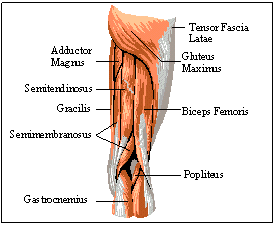
The Sartorius, Pectineus, Gluteus Medius, Gluteus Minimus, Adductor Longus, Adductor Magnus, and Adductor Brevis contribute to the rotation of the hip. Gluteus Maximus, Semimembranosus, and Semitendinosus all aid in extension and hyperextension of the hip during the stance phase. The superficial muscles can been seen in figure 5 and figure 6. (8, N/A) Other muscles that stabilize the hip are Piriformis, Obturator’s Internus and Externus, Quadratus Femoris, and the Gemellus Superior and Inferior, which are commonly known as the Gemelli Brothers. (4, 53)
 Figure 6: Posterior Muscles of the Thigh
Muscle Involvement on the Knee
The muscles that carry out the motion for the knee joint are also numerous and differ for each phase. In the first half of the swing phase, flexion of the knee joint is executed primarily by the Biceps Femoris, Semimembranosus, Semitendinosus, Sartorius, and the Popliteus. In the second half of the swing phase involves extension. This extension occurs by contraction of Vastus Lateralis, Vastus Medialis, Vastus Intermedium, and the Rectus Femoris. During the stance phase, all of these muscles are in a state of contraction to support the body’s weight.
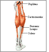
 Figure 7: Posterior Leg Muscles
Muscle Involvement on the Ankle
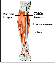
There are countless numbers of muscles that are active in the ankle and foot, but there are only four that primarily used for dorsiflexion, plantar flexion, and lateral rotation. The Tibialis Anterior is the primary dorsiflexor in the first of the swing phase. The Peroneus Longus is responsible for the lateral rotation. Both the Gastrocnemius and the Soleus contribute for the plantar flexion in the last of the swing phase. (7, 416) The muscles are illustrated in figure 7 and figure 8. (8, N/A)
 Figure 8: Anterior Leg Muscles
Conclusion
Walking is a very important and necessary process which includes three phases. There are two swing with a stance phase in between the them in each leg. The gait cycle involves numerous motions, mostly which occur in the sagittal plane, and angular displacement occurs around each joint involved with gait. The joints are located at the hip, the knee, and the ankle. This information is related to the kinematics of walking. Kinematics is the quantitative analysis or description of a motion. In the study of literature section, the mechanical principles that relate to the human gait were discussed; various malfunctions, pathologies, and abnormalities that occur in the gait cycle were explained; and the factors that influence a gait analysis, which is the study of the walking process of a patient’s by a medical professional, are introduced. Gait Analysis is used to diagnose abnormalities of the gait cycle by comparing it to the standards set by other doctors. Finally, the muscles involved in moving the lower limbs during the walking process are listed with each joint they act on, how they act on it, and in which section of the phases they act in. This paper provides informative material related to the walking process, gait analysis, and physical therapy.
Back to Physical Therapy
Back to the Brain

This page is maintained by Dickie Gipson, for comments or questions e-mail The Brain.
This page hosted by
 Get your own Free Home Page
Web Designs supplied by

Graphic Design supplied by


