The Physical Therapy Homepage
The Hand and Causes of Carpal Tunnel Syndrome
By Dickie Gipson
Technical Writing
October 18, 1996
Introduction
The hand is a part of the human body that is made up of various types of organic tissue and is used constantly to perform numerous tasks. The hand is depended upon to help people to write papers, type and do computer work, transport items, and countless other daily activities. This description is an aid for those who must use their hands to perform repetitive tasks and the people who are in charge of employees who work extensively with their hands. The description assists these people in understanding the hand, its structure, and how its parts connect to function properly. However, describing the countless number of parts in the hand is not feasible. Therefore, this description only focuses on the parts that are directly related to the condition known as Carpal Tunnel Syndrome. The description also points out what occurs in each part to cause Carpal Tunnel Syndrome.
The Parts of the Hand
The wrist bones give the hand its structure and support. The wrist includes the bones of the forearm, the radius and ulna, and the carpal bones of the wrist. Attached to these bones are the muscles. The flexors, extensors, and thenar muscles all contribute to the movement of the hand and wrist. The carpal tunnel, the third part of the hand, is the area in the wrist between the tendons and the carpals. Due to the location of the carpal tunnel, any abnormalities in this area such as swelling in muscles due to repetitive movements, can lead to numbness, and limited use of the wrist, and the hand. This condition is known as Carpal Tunnel Syndrome. The parts of the hand are illustrated in figure 1 as the wrist bones, the wrist muscles, and the carpal tunnel.
 Figure 1a: Bones of the Wrist
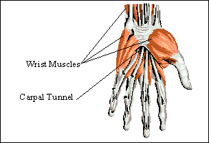 Figure 1b: Muscles and Carpal Tunnel of the Wrist
The Wrist Bones
Bones are very important structures that are made mostly of condensed calcium and a form of connective tissue. Without bones, the human body would have no structure and be more like a big blob of skin and tissue. Bones make movement possible in several ways such as providing muscles with a rigid attachment on which they can pull. Bones also make up joints which give the muscles a specific part of the body on which to act. Joints are where two or more bones meet together. For example, the wrist joint allows muscles to act specifically on the hand. The wrist joint is composed of two forearm bones, the radius and the ulna, and a group of bones collectively known as the carpals. While each of these bones have specific shapes and sizes, giving exact dimensions of the bones or any other body part is not available. This is due to the wide variations that occurs in each individual person. However, general size and shape can and has been described. The radius, ulna, and carpals are illustrated on figure 2 and covered in detail by subparts on each individual bone.
 Figure 2: The Wrist Bones
The Radius:
The radius is a long bone that is located in the forearm of the body and is a part of both the wrist and elbow joints. The radius is round and thin in the middle section known as the shaft, and contains two widened heads at the ends. This document will only be concerned with the radial head which composes the wrist joint. The radius articulates or meets the carpals of the hand on the thumb-side. The radius, due to its length, possesses numerous attachments for muscles that make up the forearm and the hand. Without the radius present all wrist joint movements would not take place. The radius also gives the hand a rigid or strong structure to enable the lifting of any object, whether it is light or heavy in weight. Without the radius to supply support, movement or simple action such as picking up a paperclip would be impossible. This would make life very difficult since the hand makes an average of one thousand movements in a single day according to Grolierís Electronic Encyclopedia.
The Ulna:
The ulna is another long bone that is found in the forearm of the body, and like its counterpart, the radius, the ulna is used for muscle attachment and supplying support to the hand. The ulna articulates with the carpals on the pinky-finger side of the hand and is the second bone that is part of the wrist. What has been said for the radius can also be said for the ulna. No movement of the hand or lifting of an object could be completed without the ulna being present in the forearm. The ulna in partnership with the radius is also able to cross over one another allowing rotation of the wrist and hand. However, the ulna is smaller in diameter than the radius and has smaller heads on the ends as well. This causes the ulna to possess smaller muscle attachments and lend less support to the hand than the radius. The ulna is still very important and what is considered normal activities of the hand could not be done without it.
The Carpals:
The carpals are a collective group of bones that connect the forearm to the hand. This group actually constitute what most people call their wrist. The carpals articulate with the radius and the ulna and provide a solid internal base of support for the hand and the fingers. If the carpals were absent from the wrist, the hand would simply be a loose hanging piece of skin that could serve no practical purpose. These bones range in shape and size and act as connectors for the hand to the forearm. The carpals provide numerous projections and ridges to allow muscle attachments for not only muscles in the hand but muscles that extend down from the forearm that are used for wrist and hand movement. Two of the carpals have projections that stick out on the palm-side of the hand.
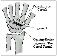 Figure 3: Details on Carpals
These two projections, labeled in figure 3, provide attachment sites for a ligament. A ligament is type of tough tissue that connects bone to bone. This ligament connects across the carpals, creating a tunnel that nerves, blood vessels, and other tissues run through. The tunnel is known as carpal tunnel and is surrounded by muscle.
The Muscles of the Wrist
Muscles are a type of tissue that has the ability to contract and relax in order to cause movement of a bone or body part. The hand is made up of thirty-three different muscles that enable it to move according to Microsoft Encarta Electronic Encyclopedia. These muscles can be divided into three basic groups. The larger muscles in the forearm are called flexors and extensors and attribute for most wrist and hand movement. However, there are smaller muscles in the hand which allow for fine motor movements. These smaller muscles are known as thenar muscles. When all three groups of muscles work together, they enable the hand to have power, grip, and explicit hand movements. The flexors, extensors, and thenar muscles have been illustrated and labeled in figure 4.
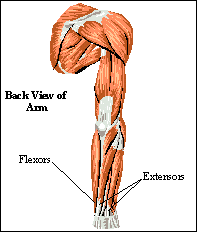 Figure 4a: Muscles of the Arm that Act on Wrist and Hand
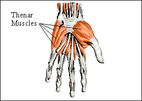 Figure 4b: Muscles of the Hand
The Flexors:
The flexor group is made up of a set of muscles located in the forearm. When placing the hand out in front with the palm of the hand facing down, the flexors can be found on the bottom side of the forearm. The flexors attach to the radius and ulna just below the elbow and extend down the arm where they attach again in various places in the hand. The flexors in the forearm are important for flexion of the wrist and hand. Flexion occurs when these muscles contract causing the wrist to bend down. For example, with the hand out in front, palm down as before, contracting of the flexor muscles causes the hand and fingers to point toward the floor. This action is called flexion and is a vital movement for the wrist, hand, and fingers in countless activities.
The Extensors:
The extensors are a group of muscles that, like the flexors, are located in the forearm. However, with the arm out and the palm down, the extensors are located on the top of the forearm just opposite to the flexor muscles. The extensors attach to the radius and ulna on the bottom side and extend down the arm where they attach to various places in the palm of the hand. The extensors cause two movements on the hand. The first, extension, causes the hand to return to a straight position after being flexed. In other words, flexing the hand caused the fingers to point toward the floor. Now take the hand and return it so that the fingers are pointing straight in front again. This action just performed is call extension. The second movement, hyperextension, occurs when the arm and hand are out in front with the palm facing down, and the wrist and hand are raised so that the fingers are pointing toward the ceiling. Extension and hyperextension, like flexion, are both very important for normal hand activities. The Extensors have tendons that run through the carpal tunnel. Extensive use of extensors can cause these tendons to swell inside the tunnel pushing the other tendons and nerves to the sides of the tunnel until they become pinched.
Thenar Muscles:
The thenar muscles are smaller muscles located in the hand. These muscles run from the thumb and the four fingers and attach around the carpal tunnel. Thenar muscles are necessary for fine movements of the hand. For example, these muscles are needed to grip a pin, to type, or to do any kind of lifting. When the thenar muscles are overused, they become swollen. This can cause Carpal Tunnel Syndrome by compressing on the tunnel and pinching the tendons and nerves in it.
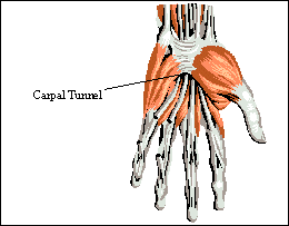 Figure 5: Carpal Tunnel
The Carpal Tunnel
The carpal tunnel, which can be seen in figure 5, is a narrow opening in the wrist in which many important nerves and tendons pass through. It is made up of eight bones in the wrist collectively known as the carpal bones and a ligament called the transverse carpal ligament. The tunnel has four walls, three of which are made up of the carpal bones themselves. The transverse carpal ligament makes up the fourth wall. A nerve, named the median nerve, passes through the carpal tunnel carrying signals between the hands and the brain. The median nerve allows the hand to move and feel sensations. Also, passing through the tunnel are nine tendons. The carpal tunnel becomes smaller if anything inside it becomes larger due to swelling or if anything that surrounds the carpal tunnel on the outside swells pressing on the tunnel itself. This can cause a pinching of the median nerve and the nine tendons encased by the carpal tunnel. The resulting injury to the hand is loss in feeling and movement. This condition, known as Carpal Tunnel Syndrome, is a serious and debilitating for the people afflicted. Carpal Tunnel Syndrome is a growing concern in todayís society due to the increase use of computers in the workplace.
Conclusion
The hand is a very important and necessary mechanism for numerous activities whether it be at the workplace or the home. This description provides the knowledge such as function of the hand, parts of the hand, the partsí functions and how each part relates to cause Carpal Tunnel Syndrome. The parts of the hand are broken down into the wrist bones, which provide structure and support to the hand; the wrist muscles, which contract and relax to cause movement of the hand; the carpal tunnel which houses and protects the median nerve and the nine tendons that pass through it. Any swelling from a muscle being overworked in or around the carpal tunnel can lead to the pinching of the median nerve and the tendons. The result is a loss of feeling and movement in the hand. This affliction is known as Carpal Tunnel Syndrome.
Back to Physical Therapy
Back to the Brain
 This page is maintained by Dickie Gipson, for comments or questions e-mail The Brain.
This page hosted by
 Get your own Free Home Page
Web Designs supplied by

Graphic Design supplied by
