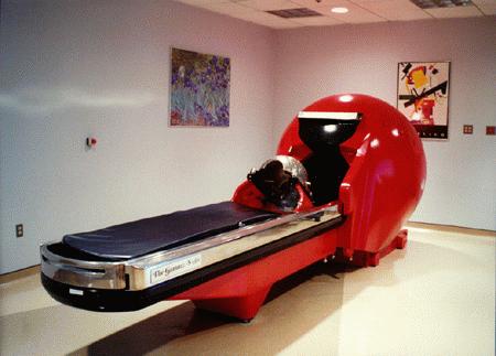
A relatively high number of inextirpable meningiomas and recurrences provides the rationale for the use of Gamma surgery in these lesions. A relative lack of published series on the results of radiosurgery for meningiomas prompted us to put our results here.
Gamma knife surgery has been performed on 220 meningiomas treated by us over the last 6 years. Imaging studies for follow up periods ranging from 1 to 6 years were available on 151 of these patients. All radiological images were analyzed with a computer using software developed by us to obtain volume measurements on the tumors. The tumors treated by us ranged in volume from 1 cc to 32 cc.
Decrease in volume was seen in 94(63%), while there was no change in size in 40 (26%) and increase in size was observed in 17 (11%) cases. The decrease in volume was more than 75% in 4 cases, 50-75% in 14 cases, 25-50% in 33 cases, 15 to 25% in 43 cases. Decrease in size of less than 15% was ignored as within the margin of error of the method. No clinical side effects or mortality was observed.
Microsurgery should be carried out in meningiomas both for extirpation or debulking and histology. The quality of life of the patient should be preserved at all costs, and where this aim cannot be fulfilled by microsurgery, radiosurgery can be carried out, as either a primary modality or in addition to microsurgery.
MRI scans of the brain following contrast administration showing a Meningioma of the cavernous sinus (A,B) before Gamma Knife surgery and 18 months later (C,D). Except for a small area of enhancement there is no evidence of residual tumor.
