(S. A. Patney: InteRyc Volume-1,Jan-Mar-2001,pp 13-23)
SHORT REVIEW ARTICLE ON STRABISMUS
CYCLOVERTICAL DEVIATIONS
Cyclovertical deviations comprise a group, which includes cases
of vertical and/or torsional (latent and manifest) strabismus. However, purely vertical
deviations are a rarity. So is the presence of true comitance in cases of vertical
deviations. Mostly they are associated with a certain amount of horizontal deviation. When
the angle is measured in 9 cardinal directions (or diagnostic positions) of gaze, a
varying degree of incomitance is usually present and on studying the results of various
tests one can frequently find some indication of the presence of a paretic factor. In many
cases though, this may be difficult to establish.
Lyle has given very pertinent quotes by Chavasse, at the start of
each chapter in his book. The one at the beginning of the chapter on vertical squint is
given here, "The cyclovertical deviations have constituted, both in pathology and
diagnosis, one of the most baffling of all problems which present themselves in the
kaleidoscopic panorama of strabismus". It is as true today as it was when Chavasse
wrote his wonderful book.
Incidence
The incidence of a vertical deviation in cases of horizontal
strabismus seems to be quite high. The various figures given in the literature are: 43% in
457 cases of convergent strabismus2, 50% in 79 cases of esotropia3,
79% in 615 cases of horizontal strabismus4 and 26% in Lyle’s series of 298
cases of concomitant convergent squint1.
Etiology
The etiology depends on the type of vertical strabismus and will be
discussed separately under each type.
Classification and symptomatology (clinical picture)
Vertical strabismus can be classified in various ways. Two of the
most commonly used classifications, one old and the other more recent, are given here, as
each is useful in a different way. Purely vertical deviations are rare. Mostly they are
accompanied by a varying degree of horizontal deviation.
Earlier classifications of vertical deviations including that
mentioned by Lyle1 and advocated by Villaseca5, divided the vertical
deviations into the following three main types:
- Primary vertical squints
due to palsy of vertically acting
muscles
- Secondary vertical squints
, as a consequence of horizontal
squints
- Mixed cases
A more recent classification is the one advocated by von Noorden6
which is as follows:
- Nonparalytic cyclovertical deviations
- Concomitant Hyperdeviations
- Dissociated vertical deviations (DVD)
- Upshoot in adduction (Strabismus sursoadductorius)
- Downshoot in adduction (Strabismus deorsoadductorius)
- Vertical deviations due to mechanical factors
- Paralytic cyclovertical deviations
Following is the description of some of the main types of
cyclovertical strabismus.
- Primary vertical strabismus (type 1 of the Vellaseca’s
classification): This group consists of cases, in which the vertical deviation
is the original one and the horizontal deviation, if present, comes later as a consequence
to disturbance of binocular fixation and fusion. There may be a latent or a manifest
deviation (intermittent or constant). It has been classified into the following 6 types:
- Primary vertical deviation
due to unilateral paresis of an
elevator or a depressor:
(A) Paresis of superior oblique or superior rectus
(B) Paresis of inferior oblique or inferior rectus
- Primary vertical deviation due to unilateral paresis of both
elevators or both depressors
- Primary vertical deviation due to bilateral paresis of the same
muscle of each eye
- Mixed or multiple paresis
- Concomitant hypertropia
- Dissociated vertical divergence (or alternating sursumduction)
- Secondary vertical strabismus occurring as a consequence of
horizontal heterotropia: A hyperdeviation is common in cases of esotropia as well as
exotropia. There is no evidence of a vertical muscle paresis, although this fact does not
rule out the possibility that vertical deviation was paretic in nature and with the
passage of time the paresis has largely recovered and the deviation has become
concomitant. The secondary vertical deviations have been divided into the following types
by Urist7:
[1] Esotropia with bilateral elevation in adduction
[2] Esotropia with bilateral depression in adduction
[3] Exotropia with bilateral elevation in adduction
[4] Exotropia with bilateral depression in adduction
- Mixed cases
are difficult to diagnose and manage. They are
uncommon. There may be various combinations as given below:
- There may be present two different types of primary vertical
strabismus in the same case, e.g., dissociated vertical divergence and vertical muscle
palsy.
- A secondary vertical deviation may occur as a consequence of
secondary horizontal deviation, which in turn was consequent upon a primary paresis of
vertically acting muscles.
- Two different types of vertical muscle paresis may occur in the same
case leading to various combinations of muscle sequels and secondary horizontal
strabismus.
Differential diagnosis of primary and secondary vertical
strabismus is of utmost importance for planning proper line of management. The
various points which help in differentiating between them (8 and 9) are enumerated in the
table 37-1:
Table 1
| |
Primary vertical deviations |
Secondary vertical
deviations |
| 1 |
The angle of squint is larger |
The angle is smaller |
| 2 |
Significant squint in PP |
Insignificant or absent |
| 3 |
Angle increases in one of the oblique
directions depending on the vertically acting muscle affected. |
Mainly present in lateroversion. |
| 4 |
Horizontal angle does not vary in PP,
elevation or depression. |
Horizontal deviation may vary increasing
in elevation or depression. |
| 5 |
A paretic vertical squint usually shows
some limitation of ocular motility. |
Function of vertically acting muscles is
normal. |
| 6 |
Bielchowsky’s sign is + in SO palsy |
The sign is negative. |
| 7 |
CHP, especially head tilt is an indication
of primary vertical squint. |
Head tilt is absent unless adopted for
esophoria or exophoria |
| 8 |
The eye with congenital ocular palsy
retains some function and vision |
Amblyopia and suppression is common in
unilateral congenital ET. |
| 9 |
A vertical deviation in the fixing eye is
usually primary. |
A secondary vertical deviation is present
in eye with horizontal squint |
| 10 |
A marked elevation in adduction indicates
primary vertical deviation. |
In secondary vertical deviation elevation
in adduction is less marked. |
| 11 |
It is more marked in oblique
gaze. |
It is more pronounced in
extreme horizontal gaze. |
| 12 |
A marked upshoot in adduction usually
means SO palsy unless proved otherwise |
The secondary upshoot in adduction is
usually mild, found in a case of long standing esotropia. |
| 13 |
Other neurological disturbances like
nystagmus and alternating hyperphoria (DVD) normally absent |
Other neurological disturbances like
nystagmus and DVD much more likely to be present. |
| 14 |
Presence of cyclodeviation likely |
Cyclodeviation normally absent |
| 15 |
Surgery on horizontal muscles does not
abolish the vertical deviation. |
Correction of horizontal squint often
abolishes he vertical deviation, if no contractures present. |
The main points regarding vertical deviations are:
- A pre-existing vertical deviation in the unoperated eye could have
been missed/masked.
- The masking of a vertical element may be explained in the following
way: A vertical deviation caused by vertical rectus dysfunction is more marked in
abduction and that caused by an oblique in adduction. Thus, a vertical element due to
oblique overaction is more likely to occur in esotropia and may be masked in exotropia.
When the exotropia without evident vertical deviation is overcorrected leading to
consecutive esotropia, the preoperative masked vertical deviation due to oblique
overaction may appear. Conversely, a masked preoperative vertical deviation due to
vertical muscle dysfunction in esotropia may appear after it has been converted into
exotropia by surgery (consecutive exotropia). The hypotropic eye is usually the fixing eye
(even if it is the affected one) as most of the daily activities like eating, walking and
reading etc. are carried out in depression.
- If the sound eye fixes and the affected eye is hypotropic, there is a
pseudo-ptosis, which must be recognized for what it is, to avoid unwanted ptosis surgery.
A simple test is all we need for differentiating between a true and pseudo-ptosis. The
hypotropic eye with the ptosis is made to fix. In the case of a pseudo-ptosis due to
hypotropia (and lid following the movement of the eye), the ptosis disappears when the eye
is placed in primary position.
- It should be remembered that the affected eye is not always the
deviating eye and that the muscle with dysfunction is not necessarily the paretic muscle.
On the contrary, it is often the ipsilateral antagonist or the contralateral synergist10.
The side of the fixing eye will determine whether it is to be the former or the latter.
- According to the first of The Four Golden Rules mentioned by
Pratt- Johnson11, in every case of vertical strabismus, superior oblique
palsy is the cause unless proved otherwise. The other three follow in the following
text.
- A paralysis of superior oblique (SO) is congenital unless proved
otherwise.
A congenital palsy may not cause symptoms until much later, usually middle
age. However, a compensatory head posture (CHP) is usually adopted to achieve binocular
single vision in straight-ahead position. Childhood photographs showing the presence of a
CHP consistent with the type of the paralysis or paresis can provide the best evidence of
a congenital palsy, in the absence of a history of double vision or squint. Sometimes
there is a history of intermittent diplopia for years. When the deviation decompensates,
the diplopia becomes more frequent or there are symptoms of strain during the use of the
eyes.
- The second commonest cause of SO palsy after congenital, is trauma,
usually a head injury.
The head injury need not be serious involving loss of
consciousness.
- If the SO palsy is not congenital (compensated or otherwise) or
traumatic, a neurological cause including an intracranial lesion must be excluded.
- It is necessary to do a full ocular motility workout in every case of
vertical deviations, even if it seems to be concomitant and secondary. Many a times the
seemingly secondary deviation turns out to be a primary one. For instance, a case of
alternating esotropia with mild bilateral upshoot in adduction would appear to be a case
of primary esotropia (especially in a child) with a secondary bilateral inferior oblique
overaction. After a full orthoptic check however, the Maddox chart (a chart showing the
angle of deviation in 9 diagnostic positions of gaze) may reveal a bilateral superior
oblique paresis indicated by an increase of the vertical component in oblique directions
and/or a presence of cyclodeviation. If it is a superior oblique palsy the vertical
deviation is maximum in contralaterodepression (depression of the eye in opposite
direction), the field of action of the superior oblique.
- Any surgery to correct the deviation must include vertical muscles if
it is a primary vertical deviation. Secondary vertical deviations do not need correction
unless there are contractures. Commonly, correction of horizontal strabismus leads to the
disappearance of the secondary vertical deviation.
- It is not an uncommon experience in a case of horizontal heterotropia
with unilateral vertical deviation (e.g., an upshoot in adduction) to have a vertical
deviation (upshoot in adduction) appear in the other eye after surgical correction.
This may be due to the following factors:
- The other view that a vertically misplaced reattachment of horizontal
muscles in the operated eye might have caused it does not seem to hold true. To cause a
significant vertical deviation the vertical displacement of the re-insertion of the
operated muscle has to be significant too12.
- It might be a case of dissociated vertical deviation (DVD) or
alternating sursumduction in which the vertical element in the unoperated eye was much
less as compared to the operated eye.
A. Nonparalytic cyclovertical deviations according to Noorden’s
classification
Each of the following four conditions will be discussed in short.
1) Concomitant hypertropia or hyperdeviations
Incidence
Truly concomitant hypertropia (hyperdeviation with the same angle in
all the directions of gaze as well as each eye fixing) is a very rare condition. Usually
there is some amount of incomitance to be found in one direction of the gaze or the other.
One must examine repeatedly to make sure.
Etiology
The cause of the small concomitant hyperdeviation which is all there
is to be found in these cases, is not known. Some of the possible explanations are
mentioned below:
- To start with, there might have been a paretic vertical strabismus,
which partially recovered and developed comitance as the time went by, as is the rule in
cases of paralytic strabismus.
- Minor anatomic anomalies may be present causing an anomalous position
of rest.
- In yet other cases abnormal innervation may be responsible.
- An abnormal position of rest may have been caused by the presence of
some minor mechanical factors.
Management of concomitant hypertropia
In many cases without symptoms or complications no treatment is
required. Sometimes when the condition is giving rise to symptoms of strain or other
problems the following modalities of therapy may be considered:
- As the degree of deviation is small and the strabismus is
concomitant, prismotherapy is usually sufficient to relieve the symptoms. The minimum
power of the prism that controls diplopia is prescribed, distributed equally
between the two eyes. The apex of the prism is placed in the direction of the vertical
deviation, which means apex up (described as base down) for hypertropia and apex down
(described as base up) for hypotropia.
- Surgery may be required in some cases to correct a co-existing
horizontal deviation. There is no need to operate on the vertical muscles. The small
amount of the vertical deviation can be managed by vertical transposition of the
horizontal muscles (while they are being recessed or resected) as follows:
- For hypertropia:
The insertions of the horizontal muscles are
shifted downwards so that a depression action is added to their horizontal action.
- For hypotropia:
The insertions of the horizontal muscles are
moved upwards to add an elevation action to their horizontal one.
2) Dissociated Vertical Deviations
Bielschowsky first published a detailed account of this interesting
and intriguing condition in 1896 although it had been known since 1894.
Definition
Dissociated vertical deviation is a name given to a special group of
cases where the main diagnostic feature is a spontaneous or precipitated (by disruption of
fusion) supraduction of either eye. It can be accompanied by excycloduction (of the
deviated eye), abduction (of the deviated eye) and nystagmus. Any or all these features
may be present.
Terminology - Alternative names
This condition has been given various names by different workers but
it should not cause confusion, as the main signs of DVD are fairly typical. It is always
the nonfixating eye that deviates upwards. When it is made to fixate the object the other
eye deviates upwards. Some of the other names are as follows:
Alternating sursumduction, alternating hyperphoria or hypertropia,
double hypertropia, occlusion hyperphoria or hypertropia, dissociated double hypertropia,
dissociated alternating hyperphoria or hypertropia, dissociated vertical divergence and
anatropia.
Most of these names do not carry the correct impression about the
clinical characteristics of the condition. The terms "hyperdeviation, hyperphoria and
hypertropia" should only be reserved for cases in which one (the hypertropic) eye
deviates up and the other (hypotropic) eye deviates downwards when it is not fixating. In
the case of DVD each eye takes turn to deviate up and it is always the nonfixating eye
that deviates up. No hypotropia can be demonstrated. As already mentioned the condition
can be unilateral or bilateral. Sometimes the hyperdeviation is manifest and constant in
one eye and latent or intermittently manifest in the other eye. This is specially so when
there is an accompanying horizontal heterotropia (esotropia or exotropia).
Etiology
The etiology of this condition is not clear but the results of
various studies including those of the upward movement of the deviating eye and the
movement of redress with the search coil method and also the studies of saccade point the
finger to an abnormal vertical vergence system. The saccades have been found to be
abnormal.
Because of the uncertainty about the causative mechanism many
theories have been put forward, none of them having gained universal acceptance. The
theories with the maximum support are the following:
- Bielschowsky’ s theory
- Spielmann’s theory
1. Bielschowsky’s theory
This theory tries to explain most of the signs observed in the cases
of DVD. According to this theory the dissociated vertical deviations are caused by
alternating and intermittent excitation of both subcortical centers that govern the
vertical divergence. To support the theory Bielschowsky presents the examples of seasaw
nystagmus and skew deviation. The main points of this theory are as follows:
- DVD results from alternating and intermittent excitation of both
subcortical centers. These centers are concerned with controlling the vertical divergence.
- In the usual heterophoria and heterotropia the innervational
association between the two eyes is maintained while in DVD it is intermittently suspended
so that certain movements of each eyes can take place independently of the other eye.
- The unilateral cases, according to him, may result when the voluntary
fixation reflex coexists with the involuntary action of one of the centers for vertical
divergence.
- The fixating eye maintains primary position because the voluntary
innervation to the depressors neutralizes the innervation to the elevators, which causes
the updeviation.
- Bielschowsky demonstrated that when a neutral filter is held in front
of one of the eyes to reduce the input to that eye progressively (by increasing the
density of the neutral filter) the innervation to the elevators of this eye is abnormally
increased. This is brought about by the increased effort to maintain fixation. When the
innervation to the elevators is increased and the eye keeps on fixating there is a
compensatory increase in the innervation to the depressors. As already mentioned, this is
known as Bielschowsky’s phenomenon. This is not the only instance of
simultaneous innervation of the antagonists, the other being seen during asymmetric
convergence.
- It is almost certain that the impulse for the DVD to occur arises in
the fixating eye. However, the site where this impulse for innervation to the elevators
starts is not known.
- It is important to understand the basic nature and the difference
between the two conditions, i.e., the DVD, which is innervational in origin and the
hyperdeviations, which are caused by anomalous position of rest. Both, Bielschowsky and
Spielmann, have stressed this point.
- Although elevation (updeviation) of the nonfixing eye is the most
common feature, excycloduction and abduction (divergence) of the nonfixing eye are common,
especially the former.
2. Spielmann’s theory
Spielmann put forward the view that DVD is caused by an imbalance
in the binocular stimulation. This view may get support by the fact that DVD is pretty
common in cases of infantile esotropia but it does not explain the occurrence of DVD in
the patients with normal binocular functions.
Spielmann has supported Bielschowsky view that the impulse for the
innervation to the elevators of the updeviating eye must originate in the fixating eye. In
fact he proved it by showing that no updeviation of either eye is to be seen if fixation
is prevented by interposing neutral filters in front of both eyes. When the filter is used
in front of one eye only the characteristic elevation of the nonfixating eye is seen. This
goes to show that in the case of DVD the fixation is the determining factor while in the
case of heterophoria (hyperphoria) it is the fusion. When the fusion is prevented or
disrupted the deviation is manifested (as the eye takes on the position of rest). As far
as DVD is concerned the fusion is not the determining factor as this condition is also
seen in the patients who do not have fusion.
Symptomatology and diagnosis
Main clinical features of this condition are as follows:
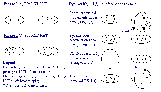
- Each eye deviates upwards [figure 1(a) and (b)] while the other eye
is fixing and this may happen under the following circumstances:
- When the patient is lost in thought or is daydreaming.
- When the patient is tired or fatigued out.
- When fusion is broken by some means like covering one eye as during a
cover test.
- When the input to the eye is reduced by interposing a neutral density
filter in front of the eye.
- Under the cover the updeviated eye may make vertical pendular
movements [figure 37-1(c)].
- When the cover is removed we see the updeviated eye moving downwards
to take up fixation in the primary position. Usually this happens spontaneously [figure
37-1(d)] when the cover is removed but sometimes this movement of redress is not
spontaneous but has to be induced by covering the other (fixing) eye [figure 37-1(e)].
- On prolonged occlusion and/or dissociation the degree of deviation
goes on increasing.
- The amount of updeviation is often asymmetrical.
- Under cover, excycloduction of the updeviated eye is common [figure
37-1(f)].
- When the cover is removed the eye is seen to cruise downwards,
incycloduct and often move medially or laterally (depending on whether there is an
accompanying divergent or convergent deviation, respectively) to take up fixation in the
primary position. The cycloduction can be detected by looking at the conjunctival blood
vessels and the iris pattern.
- Latent nystagmus
is often present in cases of dissociated
vertical deviations (DVD).
- Sometimes excycloduction of the updeviated eye is accompanied by
an incycloduction of the opposite eye. Thus if the right eye is, for the moment,
updeviated and excycloducted, there is may be seen a simultaneous incycloduction of the
left eye, the result being a cycloversion to the right (upper end of the vertical corneal
meridian in both eyes moving to the right).
- In occasional cases there is only a torsional deviation to be seen
without any vertical or horizontal elements. The excycloduction may be manifest or latent
(under cover only). During the recovery to primary position an incycloduction is seen to
occur. These cases are referred to as dissociated torsional deviation (DTD).
- In some patient one eye may have the full syndrome while the other
only the DTD.
- In some cases overaction of either the inferior oblique or the
superior oblique is found. In the latter case A pattern may be present.
- Latent nystagmus is present in 50% of cases. (NOTE: Latent nystagmus
and excycloduction were not included in Bielschowsky’s origin description of this
disease. Noorden advises use of the term "syndrome" for DVD.

|
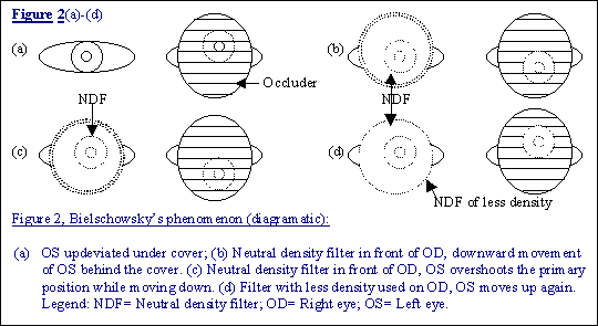
- Bielschowsky’s phenomenon: In cases of DVD when one eye
is covered, e.g., OS, it deviates upwards under the cover [figure 37-2(a)]. If, then a
neutral density filter is interposed in front of the fixing eye [OD in figure 37-2(b)]
while the other is covered and updeviated under that cover, the latter moves
downwards. It may even overshoot the mark and go down beyond the primary position mark
[figure 37-2(c)]. When the density of the neutral filter is reduced resulting in an
increase in the visual input to the fixing eye, the eye behind the cover is seen to
gradually move up again [figure 37-2(d)].
- DVD is mostly bilateral but asymmetrical.
- DVD is mostly associated with esotropia or exotropia although there
are some cases in which it is the only sign present. In such patients the binocular
functions are normal.
- In cases of infantile strabismus (esotropia and exotropia) the
incidence of DVD is high although it may not be evident under the age of 2 years or later.
Sometimes the DVD in such cases manifests for the first time after the surgery for
horizontal heterotropia has been carried out or it may take years even after the surgery.
- DVD has also been reported in association with Duane’s
retraction syndrome (15). The relationship is not clear. It may be coincidental.
- Suppression occurs commonly if the DVD manifests spontaneously. In
such cases diplopia is rare. However, use of a red filter can manifest diplopia.
- Measurement of angle of deviation
can be done by any of the
method using alternate cover test e.g., on the synoptophore or with the prism bars.
- The diplopia test using the red filter
: The red image is always
seen (by the patient) below the white one, whichever eye is fixing, indicating that it
belongs to the hypertropic eye. Each eye thus is shown to be hypertropic in turn
(alternating sursumduction). This feature coupled with an absence of vertical muscle palsy
is diagnostic of DVD. Moreover, this test can also give an idea not only of the
magnitude of the updeviation but also help in measuring it by interposing the
appropriate prisms to neutralize the separation between the two images.
Differential diagnosis
The main differentiating points are given below in table 2:
|
Bilateral inferior oblique
overaction |
Dissociated vertical
deviations |
| 1 |
Updeviation from PP, adduction
or Abduction |
Updeviation or upshot in
adduction, never in abduction |
| 2 |
V-pattern is often present |
V pattern absent |
| 3 |
SO action normal |
SO may overact |
| 4 |
A-pattern exodeviation in
downgaze may be present |
A-pattern absent unless DVD is
associated with IO overaction |
| 5 |
Pseudoparesis of contralateral
SR + |
Absent |
| 6 |
Incycloduction absent on
refixation |
Incycloduction present on
refixation |
| 7 |
Latent nystagmus absent |
Often present |
| 8 |
Bielschowsky’s phenomenon
absent |
Bielschowsky’s phenomenon
usually present |
| 9 |
Saccadic velocity of
refixation move-200-400 degrees/sec. |
Saccadic velocity of
refixation movement:10-200 degrees/sec. |
DVD has to be differentiated from hyperphoria and
hypertropia. This does not pose much difficulty as the other eye shows hypodeviation. The
main difficulty arises when there is a case with bilateral overaction of inferior oblique
muscles. These cases can be confused with those of dissociated vertical deviations (DVD)
although a little attention to pertinent points can prevent it.
(NOTE: The second and last part of this short review article on
cyclovertical deviation will be published in InteRyc volume 2, 2001)

