InteRyc 1999 volumes 3(18-25) &
4(16-26)
A AND V (ALPHABETIC) PATTERN STRABISMUS
Alphabetic pattern strabismus, of which the most common examples are A and V pattern
horizontal deviations, has gained a lot of importance during the last few decades. It
belongs to the group of incomitant horizontal deviations. The emphasis on this important
subject that has been in evidence during the last few decades is fully justified as it is
not only due to the fact that it is a common condition when one is on the lookout for it,
but also that it is much more difficult to manage than are cases of comitant horizontal
deviations. While the only effective treatment is surgery, routine surgery often fails and
special surgical procedures have to be used.
In the past, before the terms A and V syndrome and later A and V pattern strabismus were
introduced in 1956-57, several ophthalmologists noted that many cases of horizontal
heterotropia were immune to the usual surgical procedures and the rate of failures in
these cases was high. A significant number of these cases showed the presence of vertical
muscle dysfunction and all of them demonstrated a variation in the angle of the horizontal
deviation in midline vertical gaze (direct elevation or upgaze and direct depression or
downgaze). They were classified as a group with the horizontal deviations with secondary
vertical squint. However, after 1957 (see history below) the existence of this group was
acknowledged by an increasing number of ophthalmologists and dozens of papers were written
on this important topic. New strategies were devised to treat these difficult cases and
the problem of etiology of these cases is still not resolved.
I have tried to give here a short overview of a very important subject with a large amount
of literature on it. The main headings have been described in short in the following text.
Definition
In A-V pattern strabismus the angle of horizontal
strabismus varies significantly while changing the gaze from elevation to depression or
vice versa. This pattern can be seen in exotropia or esotropia. Over the years most
strabismologists have agreed upon certain criteria for diagnosis. The criteria are still
slightly variable but the following definitions are acceptable to most. In the case of
A-exotropia this variation in the angle of horizontal squint has to be more than 10 PD
when the gaze is changed from elevation to depression.
In the case of V exotropia the variation has to be more than 15 PD when the gaze is
changed from depression to elevation.
In esotropia the increase in the angle has to be relatively more (15 PD) for depression
and less for elevation (10 PD). In A-esotropia the increase in midline upgaze has to be at
least 10 PD. In V esotropia it has to at least 15 PD in midline downgaze. This is due to
the fact that an increase in convergence in looking down is normal.
History
A variation in the angle of horizontal strabismus in vertical gazes was first described by
Duane in 1897.
A-V syndromes or patterns had been described as "Horizontal strabismus with
associated vertical element" from 1896 to 1956.
Albert (1) and Costenbader (2) suggested the terms A-V patterns and A-V syndromes in 1957
and 1955 respectively.
A and V syndromes, now almost universally referred to as A and V pattern strabismus, have
attracted a lot of attraction in the last few decades resulting in better management and
results. However, the etiology in most of these cases still remains obscure.
Incidence
 |
Wide variation in the figures reported
(3) |
 |
The highest percentage reported 87.7 % |
 |
The lowest figure reported 12.5 % |
 |
Most commonly reported range 15-25 % |
 |
It is seen fairly frequently in ocular
motility clinics |
 |
The variation seems to be due to an
absence of uniform criteria of diagnosis |
 |
As regards the relative prevalence of
various types V esotropia or V exotropia have been found to be most common (the latter in
a predominantly black population). |
 |
It is most common in cases of infantile
strabismus. |
 |
It can also occur later after palsy of
either lateral recti or palsy of vertical recti or oblique muscles. |
Etiology
There are various theories but as yet there is no unanimous agreement on the etiology of
alphabetic pattern strabismus. Anomalies of various muscles and other structures have been
blamed. The names of the more popular theories and main points regarding each of them are
given in the following text.
The alphabetic pattern strabismus including A and V syndromes (the more common ones) may
be due to the following factors:
1. Secondary to horizontal muscle dysfunction: According to Urist (4) and Villaseca
(5) underaction of both lateral recti leads to a weakening of divergence on looking up,
causing A pattern esotropia and weakness of both medial recti leads to insufficiency of
convergence in depression thereby giving rise to A pattern exotropia. Conversely,
overaction of lateral recti (due to increased innervation?) can lead to V pattern
exotropia and increased innervation of medial recti to V pattern esotropia. Most people do
not support this theory but disappearance of both, vertical deviation and incomitance, in
midline vertical gaze after surgery on horizontal muscle alone (usually with vertical
transposition) does indicate some sort of dysfunction of horizontal muscles in some of
these cases.
2. Secondary to vertical recti dysfunction: Brown (6) proposed that a dysfunction
of vertical recti which have a tertiary action of adduction may cause A / V pattern. For
instance, if there is an underaction of superior recti, there may be a V pattern because
of the following factors: a) weakening of the adduction action of superior recti, b)
secondary overaction of inferior obliques, causing divergence in elevation, c) overaction
of inferior recti, causing adduction (convergence) in depression and d) secondary
underaction of superior obliques causing a weakness of their abduction action in
depression causing convergence, again contributing to the production of V pattern. This
theory does not have much support as in most cases evidence of a paresis of superior recti
is not found. But in some cases this theory may be the only one, which can explain the
etiology. It may be in these cases that the horizontal transposition of vertical recti by
surgery may be working effectively.
3. Secondary to oblique muscle dysfunction and cyclo-torsional effect: This is the
most popular theory at present. High rate of success of surgery on oblique muscles, in
eradicating A or V pattern supports this theory.
An overaction of inferior obliques (in V pattern) or superior oblique (in A pattern) is a
common finding in cases of A and V pattern strabismus. The tertiary action of the obliques
is abduction. Thus a dysfunction of these muscles can cause the alphabetic pattern in one
or more of the following ways:
A) A primary or secondary overaction of inferior obliques can cause a V pattern by
increasing its abduction action in elevation.
B) A paresis of superior obliques may lead to a weak abduction in depression causing V
pattern. This effect will be exaggerated by the consequent secondary overaction of
inferior obliques, as it will cause more divergence in elevation.
C) Conversely, weakness of inferior oblique or overaction of superior oblique may cause A
pattern strabismus by weakening abduction in elevation and strengthening it in depression.
4. Anatomical anomalies of orbits, e.g., mongoloid or antimongoloid features and
torsion of orbits: A dysfunction of oblique can occur due to orbital anomalies.
Usually A pattern esotropia with underacting inferior obliques (IO) has been reported in
patients with mongoloid features and V pattern exotropia with overacting IO in
antimongoloids. The opposite was found to be present in cases with antimongoloid features
(7). However there are cases in which the opposite is found to be true (8).
The important points to remember in this context
are:
 |
The association between the type of
features and A-V patterns is not constant. There are cases with these features are present
without A-V patterns; e.g., in many patients with mongoloid features there is no oblique
dysfunction. The reverse is also true. |
 |
Anomalies in the muscle planes and the
angle they form with the optical axes have been blamed for the oblique muscles'
dysfunction in these cases (9). Normally the obliques subtend an identical angle of 54-55
(SO) and 51 degrees (IO). Because of this slight difference, the IO is at a relative
advantage in cases of large angle esotropia which fact may explain the presence of a
secondary IO overaction in many cases. When this angle is significantly different in the
case of the two muscles, it is called desagittalization (10) of obliques. The latter may
cause an imbalance between the two muscles resulting in A, V and X patterns. |
 |
Overaction of inferior oblique muscles
is found in many cases of craniofacial dysostoses (e.g., plagiocephaly with premature
unilateral closure of coronal sutures) and hydrocephalus. |
 |
The new imaging techniques are expected
to shed new light on the etiological aspect of a great many, so called primary dysfunction
of the extra-ocular muscles, especially the obliques. |
 |
The whole orbits with their contents
have been shown to be intorted or extorted in cases of craniofacial dysostoses (11). |
 |
Intorsion of the orbits causes
intorsion of the globes and this in turn displaces the insertions of the superior recti
inwards (strengthening their adduction action in elevation) and that of inferior recti
outward (weakening their adduction action), causing A pattern and vice versa. |
5. Anomalies of muscle insertions: The main points are:
 |
Several workers have reported a
vertical displacement of medial and/or lateral rectus insertions, e.g., in V pattern
deviations, an up displacement of medial rectus, weakening its adduction action in
elevation, causing lateral rectus to abduct more and/or a down displacement of lateral
rectus weakening its abduction action in depression causing the medial rectus to adduct
more in depression. In A pattern strabismus the reverse was found to be the case (12). |
 |
Torsion of the globe (intorsion or
extorsion) can certainly cause a displacement of the insertions as described above. In
fact some workers have provided proof of the presence of torsion by various means like
fundus photography and campometry (showing a displacement of the blind spot in a vertical
direction) in cases of A and V pattern strabismus. |
 |
Loss of fusion has been blamed for
causing the cyclotorsion, as happens when the fusion is lost after an overcorrection of
exotropia. This in turn produces A or V pattern. |
Note: For a more detailed discussion on point 1-5
above, refer to reference no. 13.
6. Anomalies of muscle pulleys: In 1995 Demer
et al published a paper (14), reporting discovery of evidence for the presence of
fibromuscular pulleys of the recti and inferior oblique extraocular muscles. This paper
was one of several to follow (15-20), the last being in 1998 in which he blamed the
heterotopy of the muscle pulleys for many cases of incomitant strabismus including those
with an alphabetic pattern. The evidence that he produced was obtained by high-resolution
orbital imaging (magnetic resonance imaging or MRI) of the muscle pulleys.
Demer et al re-examined the orbital anatomy and found that the 4 recti muscles and the
inferior oblique muscle pass through connective tissue sleeves in the posterior Tenon's
fascia. These sleeves or pulleys constrain the muscle paths and thus act as the functional
origins of these muscles in the same way as the trochlear pulley does for superior oblique
tendon. These fibromuscular pulleys of the recti and the inferior oblique muscles are made
of collagen and elastin stiffened by richly innervated smooth muscle. They are anchored to
the orbital bones and to each other by means of connective tissue bands.
If these pulleys are displaced (heterotopy of the pulleys), incomitant deviations can be
caused. Demer et al found that malpositioning of rectus muscle pulleys is present in many
cases of incomitant strabismus that had been blamed on oblique muscle dysfunction. These
cases can be adequately managed by rectus muscle surgery alone.
I fully agree with their conclusion that "it is not necessary to suppose unproved
mechanisms of oblique muscle over- or underaction".
Symptomatology and clinical picture
Generally speaking, V or Y pattern strabismus with orthophoria/latent deviation in PP
and/or in depression produces little or no symptoms.
A-pattern strabismus may cause diplopia and/or asthenopia
We have found that the most common complaint is an intermittent squint with suppression.
The symptoms are usually absent except in the patients with a decompensated heterophoria
in depression when asthenopia may be present.
The usual complaint in this group is a deviation manifesting either in certain directions
(e.g., in elevation) or for distance, which causes a cosmetic problem. Thus V pattern
exotropia cases come complaining of a cosmetic problem more often than others do as their
deviation manifests when they look up.
Investigations
The usual investigations routine to any case having problems of ocular motility and
binocular vision are carried out in these cases also, e.g., cover test, Bielchowsky's head
tilting test, examination of ocular movements, convergence, visual acuity, refraction
under cycloplegia, fundus examination, tests for the presence of binocular functions, and
measurement of deviation. In addition, just like any other case of strabismus, any other
special tests have to be carried out if indicated, e.g., Hess chart and diplopia test if
there is a suspicion of the presence of palsy.
In these cases the angle of deviation should be measured fixing each eye in turn (to
reveal any paretic component) in at least 3 vertical directions as follows:
1. In 30 degrees elevation
2. In primary position (PP)
3. In 30 degrees depression
Note: Measurement in these three directions is all that is needed if vertical and oblique
movements appear to be normal. If however, paresis is suspected it is better to get
measurement in all the 9 cardinal directions of gaze.
Many ophthalmologists advocate a 25 degrees movement of the fixing eye from the PP, other
even less. In fact there is no standard as yet fixed in this respect. I find 30 degrees
excursions quite adequate.
Important points regarding the investigations of the cases of A and V pattern strabismus:
 |
One should especially look out for the
presence of a compensatory head posture (CHP) which usually means that either paretic or
an alphabetic pattern strabismus is present. |
 |
Patient should be wearing full
correction otherwise the information obtained may be wrong. |
 |
Measurements should be taken both
before & after surgery. |
 |
They should be taken for distance
wearing full refractive correction. |
 |
Synoptophore or prism bars can be used
for measuring the deviation. |
 |
To ensure exact degree of movement of
fixing eye from PP, I prefer using a synoptophore because the degree of movement of the
fixing eye can be accurately controlled, because it is possible to measure the torsional
deviations as well as the vertical and horizontal ones and because the synoptophore
measures the deviation for distance as is required in these cases |
 |
If the prisms are used for measurement
the fixation object should be an accommodative object and not a light and should be placed
at 6 meters. |
 |
Ocular motility should be examined
carefully so as not to miss overaction of superior or inferior oblique muscles which is
not infrequently present in A and V pattern strabismus (respectively). |
 |
Once the measurements are taken and
recorded as follows, one has to find out if there is any significant difference between
the angle of horizontal deviation when the patient is looking 30 degrees up and when the
patient is looking 30 degrees down. |
|
|
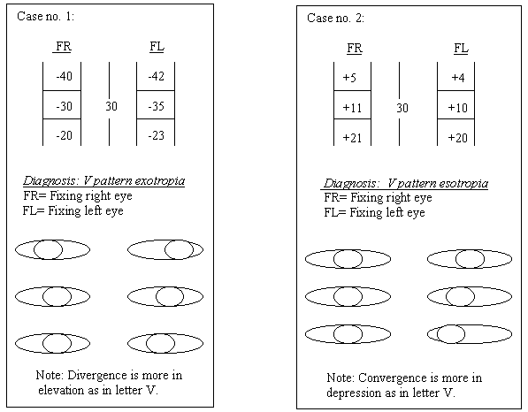
| Examples of measurements in
A and V pattern strabismus are given below: |
 |
Thus we see in the above examples the V pattern can
be present in exotropia or esotropia. Similarly A pattern can be present in either
exotropia or esotropia.
In X pattern there is orthophoria in primary position (PP) and divergence in elevation and
depression.
In Y pattern there is orthophoria in PP and depression and divergence in elevation.
In /l pattern (lambda pattern) there is orthophoria in elevation and divergence in PP and
depression.
In diamond pattern there is more divergence in PP and less divergence or orthophoria in
elevation and depression in cases of exotropia. In cases of esotropia there is more
convergence in elevation and depression and less convergence or orthophoria in PP.
The same thing can be expressed in another way, that esotropia and exotropia both can be
associated with either A or V pattern.
General principles of management
1. There are two aspects of every strabismus case: Sensory and motor. Both should be
looked after to ensure success.
2. Full correction of refractive error unless indicated otherwise
3. No surgery unless there is a manifest deviation more often than occasionally.
3. Remove suppression and treat amblyopia before surgery.
4. Always take good postoperative care
Management of A and V pattern strabismus
Full correction of refractive error
Treatment of amblyopia in children and in others with good capacity for fusion
Remove suppression in cases with intermittent strabismus and good binocular functions
Surgery if the deviation causes symptoms, cosmetic defect and/or produces sensory
anomalies like suppression/amblyopia
Indications of surgery
1. Deviation manifesting 50 % of time or more and/or
2. Presence of sensory problems, e.g., suppression, defective binocular functions
3. When it poses a cosmetic problem
4. Requirement of normal binocular functions and ocular motility for certain professions
Choice of surgical procedures
(1) Horizontal muscle surgery with vertical displacement of insertions
(2) Surgery on vertical recti with horizontal displacement of insertions, nasally or
temporally
(3) Oblique muscle surgery, in cases of significant oblique muscle dysfunction, e.g.,
inferior oblique overaction in V-pattern and superior oblique overaction in A-pattern.
Each alternative has got its proponents. Some surgeons prefer the vertical muscle surgery
as a primary procedure, others do it only if the vertical element significant. Yet others
mainly tackle the horizontal muscles unless the vertical deviation is significant and
seems to be primary in nature (and the horizontal deviation secondary to the vertical
element).
Following is a short description of the most commonly used procedures, as a detailed
description of surgical procedures is out of scope of this review. For more information
regarding surgery for Alphabetic pattern strabismus one can consult any of the strabismus
surgery books. A comprehensive discussion is given in the 5th edition (1996) edition of
von Noorden's book (15).
(1) Horizontal muscle surgery with vertical transpositioning of their insertions
This procedure has been reported to be quite effective in cases that are without
significant dysfunction of vertical muscles, especially the obliques (inferior and
superior oblique muscles). The basic principle is the fact that action of a muscle is
weakened in the direction in which its insertion is shifted, for instance, if the
insertion of lateral rectus is moved downwards, its action, that is abduction will be
weakened in depression (downgaze). The action of a medial rectus will be weakened in
elevation if its insertion is shifted upwards.
Thus, in cases of V pattern strabismus the lateral rectus insertion is moved upward and
medial rectus insertion downward. Acting on the same lines, in A pattern strabismus the
insertion of lateral rectus is moved down and that of medial rectus upwards (see the
figure below).
The amount of shift depends on the difference in the angle of deviation in midline
vertical gaze, that is to say, the increase in the angle of horizontal deviation when the
gaze is shifted from downgaze to upgaze and vice versa. Usually in a moderate case, ½
length of the insertion is moved.
We have found that many a times even significant vertical deviations have gone away after
horizontal muscle surgery. One wonders if these are the cases in which the causative
factor lies in the muscle pulleys or in the insertions of the horizontal muscles (see
discussion on etiology points no. 5 and 6.
Note: In cases of a significant amount of horizontal deviation in primary position: The
horizontal muscles are recessed or resected according to the requirement, in addition to
the vertical shift of the insertions,
(2) Surgery on oblique muscles
It is greatly favored by most surgeons, as it is very effective in reducing the A-V
patterns. In cases V pattern strabismus a weakening of overacting inferior oblique
muscles, in addition to recession-resection (as indicated) of horizontal muscles, is
indicated.
A pattern strabismus, the superior obliques have to be weakened if they are overacting.
This procedure is, of course, in addition to recession-resection of horizontal muscles as
indicated.
(3) Vertical rectus muscle surgery with horizontal transposition of their
insertions:
For A esotropia a transposition of the superior rectus muscle by 7 mm temporally increases
its abductive action and decreases the elevation action in up gaze.
Planning the line of management of pattern strabismus:
General outline of management of pattern strabismus has already been given in I part of
this article. However, certain important points have to be underlined and stressed. This
is a difficult condition to treat and thorough examination and careful planning is
essential. The following points must be kept in mind while planning surgery to decide the
muscles to be operated on and the choice of the procedure.
The points given below must be considered carefully, keeping in mind the possible side
effects or complications as a result of a certain technique. These precautions are
necessary in cases of pattern strabismus, especially A and V patterns because of not only
the complicated nature of the strabismus but also because of the complicated nature of the
actions of the extraocular muscles. The later is particularly true of oblique muscles that
have complex movements especially in extreme side gaze.
 |
Presence of a ptosis even a mild one
should be detected and its nature decided, i.e., if it is true ptosis or false. If it is a
pseudo-ptosis it is seen in the hypotropic eye while the other eye is fixing. When the
hypotropic eye with the pseudo-ptosis is made to fixate the ptosis disappears. |
 |
Exact measurements of the pattern (the
change in the angle of deviation) from direct elevation (25-30 degrees) to direct
depression (25-30 degrees) must be taken. This can be done on the major amblyoscope or
with prism bars. |
 |
Actions of the extraocular muscles
should be investigated thoroughly through examination of ocular motility so that an
overaction or underaction of a muscle is detected. Success of surgery may depend on this.
Unless these problems are corrected response to treatment may not be as anticipated |
 |
The results of the surgery may also be
unpredictable because of anomalies of the muscle pulleys and abnormalities of the orbits. |
 |
Oblique muscles should only be operated
on if they show a significant degree of dysfunction (underaction or overaction). |
 |
Vertical muscles should only be
selected for surgery if other options like surgery on horizontal or oblique muscles have
failed or are contraindicated. |
 |
In the absence of significant
dysfunction of oblique muscles, surgery should be confined to the horizontal recti
(vertical displacement with recession / resection according to the need). The action of a
rectus is weakened in the direction it's insertion is transposed, e.g., if the insertion
of medial rectus is displaced downwards as is required in V pattern strabismus, adduction
is weakened in depression |
 |
Medial rectus insertion is always
transposed towards the apex of A or V to abolish the convergence of the arms of the
letters. |
 |
Lateral rectus insertion is always
displaced towards the base of A or V to straighten the divergent arms of the letters. |
 |
Vertical transposition of the insertion
of the horizontal muscles is the only thing required if there is no manifest horizontal
deviation in primary position. |
 |
If there is a horizontal heterotropia
in primary position recession and / or resection surgery is combined with vertical
transposition of the horizontal muscles. |
 |
If there is a bilateral dysfunction of
the oblique muscles, any asymmetry in dysfunction should be noted. |
 |
As regards the surgery on oblique
muscles it should be kept in mind that weakening of an overacting oblique muscle gives
better results than does strengthening of an underacting oblique muscle. |
 |
While planning surgery of multiple
muscles specially recti one should remember that there is a possibility of anterior
segment ischemia. Oblique muscles do not contribute to the circulation of the anterior
segment. |
 |
Moderate degrees of A or V patterns
require only symmetrical surgery on the obliques or surgery on horizontal recti with
vertical repositioning of their insertions as mentioned above. |
 |
If the degree of A or V pattern is more
than 30 PD 4-6 muscles may have to be operated on but it is wiser not to do too much at
one sitting as there are so many factors affecting the outcome. And a significant
overcorrection leading to diplopia is quite an uncomfortable condition. |
 |
As far as possible both horizontal and
vertical deviations should be corrected in one sitting. |
Aims of surgery
In part I of this article indications of surgery have already been discussed. Now we take
up the aims of surgery being planned.
It is important to realize that a perfect alignment in all positions of gaze is even more
difficult in cases of pattern strabismus. One has to be realistic in approaching the
problem. The aims for surgery for pattern strabismus from a practical and realistic point
of view are three-fold:
(a) To achieve orthotropia in more practical and useful positions of gaze, i.e., in
primary position and infraversion (looking directly downwards). The latter is the position
in which most of the daily activities like walking, eating, reading and writing etc. are
performed.
(b) To obtain comfortable binocular single vision (also in primary position and looking
down).
(c) To correct abnormal (compensatory) head posture. This will automatically result if (a)
and (b) have been achieved.
Choice of the technique for various types of pattern strabismus (table 1)
Table 1, choice of technique for various types of pattern strabismus:
Type of Deviation |
Type of Pattern |
Oblique Function |
Most likely CHP |
Surgery on Extraoc. Muscle: |
Procedure |
| Esotropia |
V |
Normal BIO
+ |
Chin Ditto |
MR BIO+MR |
Recess+infraplace  Weakening |
| A |
Normal BSO
+ |
Chin Ditto |
MR BSOMR |
Recess+supraplace Weakening |
| Y |
BIO+ BSO
- |
Chin Ditto |
BIO+MR BSOMR |
Weakening Tuck BSO*,recess MR |
| Exotropia |
V |
Normal BIO+ |
Chin Ditto |
BLR BIO+LR |
Recess+supraplace Weakening |
| A |
Normal
BSO + |
Chin Ditto |
BLR BSOLR |
Recess+infraplace Weakening |
| Y |
BIO + BSO
– |
Chin  Ditto |
BIO BSO |
Weakening Strengthen (Tuck)* |
| Lambda- XT |
/\ |
Normal
BSO + |
Chin Ditto |
BLR BSO |
Infraplace Weakening |
Diamond-
XT** |
V |
Both obliques - BLR–( ) |
None(?) |
BLR ** |
Recess ** |
Legend of the table 1:
BLR: both lateral recti, BMR: both medial recti, BSO: both superior obliques,
BIO: both inferior obliques, XT: exotropia.
*: Strengthening of superior oblique is done by tucking of the SO tendon but there is a
risk of postoperative SO overaction. If too much tucking is done the tendon is shortened
and a syndrome resembling Brown's syndrome results (acquired Brown's syndrome) with a hard
and short SO tendon and positive Forced Duction Test.
**: Diamond shaped pattern is very rare. There is relative convergence of the visual axes
as compared to the angle of deviation in primary position. I have seen it a few times
associated with XT, mostly as a postoperative phenomenon. Surgery: If there is exotropia
in primary position and the lateral recti are overacting they can be recessed. If on the
other hand there is significant underaction of obliques it should be corrected.
NOTE: In cases of unilateral heterotropia with amblyopia unilateral surgery is performed.
For a significant degree of unilateral exotropia with V pattern lateral rectus recession
and supraplacement of its insertion by ½ insertion length (about 5 mm) + resection of
medial rectus and its infraplacement (insertion transposed downwards by ½ insertion
length, by about 5 mm) is done on the affected side.
Variations of the various procedures mentioned earlier have been advised.
They are used in special circumstances. The more popular ones are given below:
1. Horizontal muscle transposition:
Usually the insertion of the horizontal muscle is transposed by ½ length (about 5 mm).
However, if there is a large A or V pattern, additional effect can be obtained by
transposing the insertion by a longer length or by slanting the new insertion so that one
edge of the insertion is nearer to the limbus than the other. A few examples are given in
the following text:
A. For large patterns (more than 20 PD):
(a) Transposition of the insertion of the horizontal recti by more than ½ length
of the insertion, if the size of the pattern (the difference between the angle of
horizontal heterotropia in up-gaze and down-gaze) is large (more than 20 PD). However,
recently a study has reported that the amount of shift more than ½ length does not
correlate with the degree of postoperative correction (16). The size of the pattern
relates better with the amount of correction resulting from surgery. In addition to the
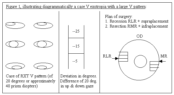
transposition, the horizontal recti are recessed or resected as the need be (figure 1) if
there is a heterotropia in primary position.
(b) Slanting the newly transposed muscle insertion for an added effect in case of
a larger pattern so that selective weakening can be achieved in elevation or depression as
required. To do this the new insertion has to be slanted.
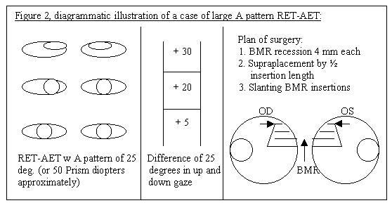
Take for example a case of A esotropia. The medial rectus has to be weakened more in
up-gaze than in down-gaze to abolish the convergence of the arms of A. This can be
achieved by transposing its insertion upwards and slanting the new insertion so that its
upper end is further away from limbus than the lower end. Of course, recession of medial
rectus is done simultaneously to abolish the esotropia in primary position also (figure
2). Putting the upper end further back further weakens the adduction action of the medial
rectus in elevation.
B. For moderate sized patterns (15-20 PD):
To weaken the action of a horizontal muscle in elevation (up gaze) selectively, the muscle
is supraplaced or transposed upwards (the group a below) or the end of its upper border is
recessed more than the end of its lower border (slanting the insertion as in group b
below).
To weaken the action of a horizontal muscle in depression (down gaze) its insertion is
infraplaced or transposed downwards (the group a below) or its new insertion is slanted so
that the end of the lower border is recessed more than the end of the upper border.
Thus there are four options as detailed below.
a) For pattern strabismus with heterotropia in primary position: Recession/
resection of the horizontal rectus muscle/muscles + vertical transposition of the new
insertion.
b) For pattern strabismus with horizontal heterotropia in primary position:
Recession/resection of horizontal rectus muscle/muscles + slanting of the recessed or
resected muscle.
c) For pattern strabismus without horizontal heterotropia in primary position:
One of the two procedures for selective weakening of a horizontal muscle (vertical
transposition or slanting its insertion) without recession/resection.
C. For small patterns (10-15 PD):
1) Without horizontal heterotropia in primary position and without a compensatory head
posture and symptoms like diplopia: no surgery is required.
2) With horizontal heterotropia without complaints surgery to correct the horizontal
deviation alone is done.
3) Without horizontal heterotropia but with symptoms in upgaze or downgaze, only the
slanting of the insertion can correct A or B pattern. The usual amount of the selective
shift of the tendon to slant its insertion is 2-mm backward (17).
2. Oblique muscle surgery
NOTE: It should never be undertaken unless there is overaction of the oblique under
consideration for surgery. This is a sound policy in order to prevent postoperative palsy
of the operated oblique muscle leading to iatrogenic vertical and torsional strabismus and
diplopia. The situation is compounded by a postoperative overaction of the direct
antagonist of the operated oblique muscle (e.g., overaction of the IO in the case of a
weakening procedure on SO).
(A) Inferior oblique muscle surgery
It is indicated if there is significant overaction of inferior obliques (IO) giving rise
to V pattern. Any of the weakening procedures given below can be carried out and the
choice depends on the individual surgeon's preference. The usual resulting correction
after any of the procedures listed below is about 20 PD in elevation (18). The effect of
bilateral IO myectomies is an esotropia of about 20 PD in elevation and no effect in
primary position and depression (19). There is no significant effect on the horizontal
alignment in primary position (19 and 20).
(1) Generally bilateral IO myectomy is quite effective.
(2) Recession of IO insertion is another good procedure.
(3) Anterior transposition of the IO has recently become popular (21).
(B) Superior oblique muscle surgery
One should be more cautious where SO surgery is concerned. Therefore weakening of the SO
should only be done in the patients with a marked degree of SO overaction producing A
pattern. The following procedures are being performed for weakening the SO:
1. SO tenotomy is the most popular technique. The main considerations in relation to SO
tenotomy are the following:
 |
There are disadvantages including more
chances of postoperative (vertical and torsional) diplopia in depression (sometimes in
primary position also), especially after bilateral tenotomy. |
 |
The postoperative result regarding
vertical alignment in primary position is rather unpredictable. |
 |
The effect of a SO tenotomy nasal to
the superior rectus (SR) tendon is more marked than that performed temporal to the SR
tendon |
 |
The effect of this procedure also
depends on the size of the pattern, larger the size more the effect. |
 |
The effect of this procedure on A
pattern (which is produced by SO overaction), in depression varies from 25-45 if done
nasal to the SR tendon (22 and 23). |
 |
The effect of SO tenotomy done temporal
to the SR tendon averages 30 PD but may correct up to 40 PD of the A pattern (18). |
 |
A graded weakening effect can be
achieved by using different techniques as discussed below*. |
 |
The effect of SO tenotomy on horizontal
alignment: SO tenotomy done nasal to the SR tendon causes a convergence of the eye by
about 3.3 PD. Done temporal to the SR tendon, the SO tenotomy results in a convergence or
a shift towards esodeviation by about 8 PD. Knowing this in advance one can plan surgery
of any associated horizontal heterotropia better. |
 |
I agree with strabismologists who
advise caution in deciding upon and performing SO tenotomy in the patients who have fusion
(17 and 24). A diplopia in depression is unbearable as most of daily activities are
carried out with eye looking down (eating, walking, reading, writing etc.). |
Choice of techniques for a weakening of the SO
muscle*:
| (1) |
Total tenotomy, nasal or temporal to SR
tendon, as mentioned above. One can try to grade the weakening effect by choosing the
procedure. The nasal tenotomy is supposed to produce more effect than does the temporal
tenotomy |
| (2) |
Cutting the attachments of the SO
tendon to the undersurface of the SR tendon can enhance the effect of total tenotomy of
the SO tendon. |
| (3) |
Partial tenectomy of the posterior
fibres of the SO tendon will not affect torsion in primary position. This procedure can be
used as an alternative to tenotomy. Resection of a segment of both SO tendons with their
sheaths can correct up to 25-35 PD of horizontal deviation in downgaze |
| (4) |
Recession of both SO tendons can also
serve the purpose (25) |
| (5) |
Desagittalization of the SO has been
advised in cases of A pattern strabismus (26). This is to widen the angle between the
visual axis and the SO tendon. |
| (6) |
A tenotomy of the posterior fibres of
the SO tendon can also reduce the A pattern in moderate cases (27). |
NOTE: Comparative studies on the relative merits and
demerits of the above mentioned procedures to weaken the SO are not available.
Strengthening of the superior oblique
Underacting superior obliques can be strengthened by a bilateral advancement of their
tendons.
This procedure is more popular in Europe. Its aim is to get rid of the convergence of the
arms of V in the patients not having significant horizontal deviation in primary position.
Complications of surgery for pattern strabismus
| 1. |
Iatrogenic (postoperative) vertical and
torsional strabismus: This complication usually results when a normally functionally
oblique muscle is weakened. |
| 2. |
Vertical and / or torsional diplopia is
a result of 1. |
| 3. |
Postoperative superior oblique palsy
after weakening of the muscle. This is also related to 1 and 2. |
| 4. |
Consecutive overaction of the direct
antagonist of the operated muscle, e.g., IO overaction resulting in V pattern after SO
weakening for A pattern. |
| 5. |
Compensatory head posture may be
produced due to the conditions mentioned under the points 1-4. |
| 6. |
Anterior segment ischemia both vertical
recti are operated in one sitting after failed horizontal rectus surgery or as a first
procedure. |
| 7. |
Persistence of oblique muscle
overaction. This complication is usually a result of a failure to diagnose or treat the
oblique muscle dysfunction. |
Summary and conclusions
 |
Treatment of pattern strabismus is more
difficult than that of comitant horizontal deviations. |
 |
The aim of treatment is to reduce the
pattern and to achieve fusion in primary position and downgaze. This will get rid of the
compensatory head posture also. If the surgery is carried out for cosmetic reasons as in V
exotropia, one should strive to align the visual axes in upgaze also. |
 |
Consider thoroughly if it is necessary
to treat the pattern. |
 |
Surgery should only be considered if
there are definite and strong reasons for treating the pattern. |
 |
The results of surgery for pattern
strabismus are rather unpredictable as in the case of vertical transposition of horizontal
recti. This is especially true if combined surgery (on recti and obliques) is carried out. |
 |
Planning the management of pattern
strabismus should be very careful and should involve thorough testing particularly for
measuring the angle of deviation in 25-30 degrees elevation (upgaze) and depression
(downgaze). One should specially look for the state of oblique muscle function and the
presence and strength of fusion. |
 |
Every patient must be evaluated
individually. |
 |
There is more chance of diplopia if the
condition is treated after 8 years of age. |
 |
However, if the surgeon is competent
and proper planning has been done the results are still worthwhile. |
X pattern
Though mentioned earlier also it must be stressed that X pattern is rather different from
other types of pattern strabismus as it does not need and usually does not respond to
treatment.
Sometimes X pattern is due to tight lateral rectus muscles (congenital or postoperative).
This condition is called "Tight Lateral Rectus Syndrome".
No treatment is required as there is orthophoria in primary position and the condition
does not usually respond to treatment. Sometimes slackening the tight lateral recti may
help.
References
| 1. |
Albert, D.G.: Personal Communication.
In Parks, M.M.: Annual Review: Strabismus, Arch. Ophthalmol. 58:152, 1957 |
| 2. |
Costenbader, F.D.: Introduction , In
Symposium: the A and V pattern in strabismus, Trans. Am. Acad. Ophthalmol. Otolaryngol.
68:354, 1964. |
| 3. |
Noorden, G.K. von: Binocular Vision and
Ocular Motility: Theory and Management of Strabismus, 5th edition, St. Louis, Mosby-Year
Book, Inc. 1996, p.382 |
| 4. |
Urist, M.J.: The etiology of the so
called A and V syndromes, Am. J. Ophthalmol. 46:835, 1958. |
| 5. |
Villaseca, A.: The A and V syndromes,
Am. J. Ophthalmol. 52:172, 1961. |
| 6. |
Brown, H.W: Vertical deviations. In
Strabismus Symposium, Trans. Am. Acad. Ophthalmol. Otolaryngol. 57:157, 1953 |
| 7. |
Noorden, von G.K.: same as in ref. No.
3, p. 379. |
| 8. |
Noorden, von G.K.: same as ref. No. 3,
p. 379. |
| 9. |
Fink, W.: The role of developmental
anomalies in vertical muscle deficits, Am. J. Ophthalmol. 40:529, 1955 |
| 10. |
Gobin, M.H.: Sagittalization of the
oblique muscles as possible cause for the "A", "V" and "X"
phenomena, Br. J. Ophthalmol. 52:13, 1968. |
| 11. |
Lemon de Brown et al: Strabismus in
plagiocephaly, J. Pediatr. Ophthalmol. Strabismus 25:180, 1988. |
| 12. |
Nakamura, T. et al: Insertion anomalies
of the horizontal muscles and dysfunction of the oblique muscles in the A-V patterns, Acta
Soc. Ophthalmol. Jpn. 95:698, 1991. |
| 13. |
Noorden, von G.K.: same as in ref. No.
3, P. 376-382. |
| 14. |
Demer, J.L. et al: Evidence for
fibromuscular pulleys of the recti extraocular muscles. Invest. Ophthalmol. Vis. Sci.
36:1125-1136, 1995. |
| 15. |
Noorden, von G.K.: same as in ref. No.
3, P. 385-389. |
| 16. |
Brooks, S.E. et al: Vertical shift of
the medial rectus muscles in the treatment of A pattern esotropia: analysis of outcome. In
Lennerstrand, G.: Update on Strabismus and Pediatric Ophthalmology, Boca Raton, Florida,
CRC Press, 1995, p. 256. |
| 17. |
Pratt-Johnson, J. and Tillson, G.:
Management of Strabismus and Amblyopia-A practical Guide, New York, Thieme Medical
Publishers, Inc., 1994, p. 141. |
| 18. |
Helveston, E.M.: A logical scheme for
the planning of strabismus surgery. In: Surgical Management of Strabismus, An Atlas of
Strabismus Surgery, St. Louis, C.V. Mosby Company, 1993, 381. |
| 19. |
Pratt-Johnson, J.: same as in ref. No.
17, p.140. |
| 20. |
Stager, D.R. and Parks, M.M.: Inferior
oblique weakening procedures: Effect on primary position horizontal alignment, Arch.
Ophthalmol. 90:15, 1973. |
| 21. |
Stager, D.R. et al: Anterior
transposition of the Inferior Oblique, Arch. Ophthalmol. 110:360-362, 1992. |
| 22. |
Parks, M.M.: Comitant vertical
deviation. In: Ocular Motility and Strabismus, 1975, Hagerstown, MD, Harper & Row, p.
133. |
| 23. |
Parks, M.M. and Mitchell, P.R.: A and V
patterns. In Tasman, W. and Jaeger, E.A., editors, Duane's Clinical Ophthalmology, Vol. 1,
Philadelphia, PA, Lippincott-Raven, 1996, p.1. |
| 24. |
Harley, R.D. and Manley, D.R.:
Bilateral superior oblique tenotomy in A pattern exotropia, Trans. Am. Ophthalmol. Soc.
67:324, 1969. |
| 25. |
Caldeira, J.A.: Bilateral recession of
the superior oblique in A pattern tropia, J. Pediatr. Ophthalmol. Strabismus 15:306, 1978. |
| 26. |
Noorden, G.K.: same as in ref.no.3, p.
387. |
| 27. |
Prieto-Diaz, J.: Management of superior
oblique overaction in A-pattern deviations, Graefe's Arch. Clin. Exp. Ophthalmol. 226:126,
1988. |


