Benign gastric ulcer

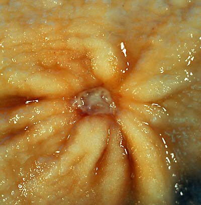
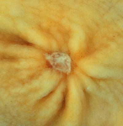
This 1-cm benign gastric antral ulcer was discovered serendipitously in a gastrectomy specimen removed for adenocarcinoma of the fundus (not shown in the photo). The gross appearance is classic for a benign ulcer in that 1) it is relatively small, 2) the mucosa surrounding the ulcer base does not appear tumefactive, and 3) the radiating rugal folds extend nearly all the way to the margins of the base. Contrast this appearance with that of the malignant gastric ulcer included in this case collection. The criteria for grossly and endoscopically distinguishing benign ulcers from cancer are not absolute, which is why it is necessary to perform a biopsy on any non-healing gastric ulcer. Even biopsies are not 100% accurate in picking up a cancer, so negative pathology reports in such cases may provide false reassurance.
The photo above was taken with a Minolta X-370 with 100mm bellows lens on Kodak Elite ISO 100 film. The specimen was previously fixed overnight in formalin after being pinned out in a wax-bottomed tray. The photo below is of the same specimen submerged in tapwater. The latter technique removes the glare in the highlights, but I prefer the sense of depth afforded by the conventional "dry" technique, above.

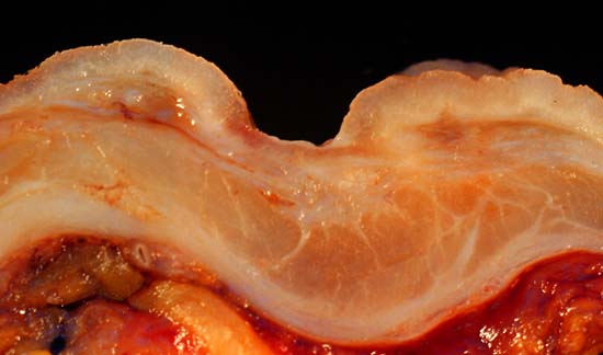
The photo below is a of a longitudinal section of the ulcer and adjacent gastric wall. Note that the normal anatomic layers are discrete and relatively undisturbed as compared with the homogenation of the wall and caused by some malignant ulcers.

Photograph by Ed Uthman, MD. Public domain. Posted 23 Sep 00
[Back to image table of contents]
[To Ed Uthman's Home Site]
for more original
resources in pathology and laboratory medicine