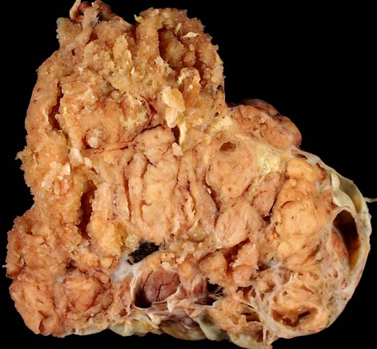
Serous Cystadenocarcinoma of the Ovary

This 17-centimeter serous cystadenocarcinoma was discovered during exploratory laparotomy of a woman who presented with intestinal obstruction, which was caused by extrinisic compression of the bowel by one of the many intra-abdominal metastases of this tumor. Grossly, the tumor's cut surface demonstrates both cystic and papillary architectural patterns.
The photo was taken with an Olympus D-600L digital camera on a copy stand illuminated by 4 photofloods. The specimen had been fixed in formalin overnight and immersed in 70% ethanol shortly before it was photographed.
Photograph by Ed Uthman, MD. Public domain. Posted 15 Jan 99
[Back to image table of contents]
[To Ed Uthman's Home Site]
for more original
resources in pathology and laboratory medicine