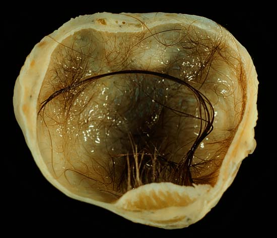
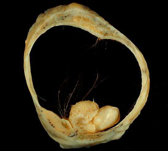
Mature cystic teratoma of ovary

This 4-cm mature cystic teratoma of the ovary was found incidentally at the time of Caesarean section and removed. Like most ovarian teratomas (and unlike those of the testis) this one was benign, showing only mature tissues microscopically. In addition to the obvious cutaneous structures (giving rise to the hair seen here), histologically there was abundant neuroglia and even one neuron. Central nervous system tissue is very common in mature teratomas.
The photo below shows a well-developed tooth arising from the right side of the mural nodule ("Rokitansky nodule") that contains most of the solid teratomatous elements. The central portion of the nodule contains mostly cutaneous tissues (skin, sweat glands, and hair follicles), while the neural tissues extend into the wall toward the left.

In the past, the teratoma (literally, "monster tumor") was considered to be a conceptus, and (according to an ancient Merck Manual I remember reading) the Roman Catholic Church required that teratomas be baptized (giving rise to interesting speculation about what the Teratoma Department in Heaven was like and who was in charge there--Saint Humpty Dumpty, perhaps?) Currently, neither pathologists nor the Vatican considers teratomas to be anything other than germ cell tumors of host origin only, with no paternal contribution.
These photos were shot on Ektachrome Elite 100 film, daylight type, through a blue filter to correct the emission spectrum of the Photofloods used on the copy stand. The exposure was f/8 for 1/4 second, using a Minolta X-370 fitted with bellows.
The key to a good photo of a teratoma is to clean out all the greasy material in the cyst. To do this, on receipt of the specimen, I palpated the external surface to determine where the mural nodule was. Then, at the antipodes of the nodule, I carefully made a straight gentle incision that extended for about half the circumference of the specimen. I did not express the contents not distort the cyst in any way. Then, I soaked the specimen in Carnoys' fluid overnight. By the next day, the grease had been broken up and diluted enough that I could gently wash it out of the lumen with more Carnoy's. This left a squeaky-clean internal surface, and all I had to do was extend my original incision all the way around the specimen, using scissors for the thin part of the wall.
Photograph by Ed Uthman, MD. Public domain. Posted 27 May 01
[Back to image table of contents]
[To Ed Uthman's Home Site]
for more original
resources in pathology and laboratory medicine