Macroadenoma (Prolactinoma)
Images and Features
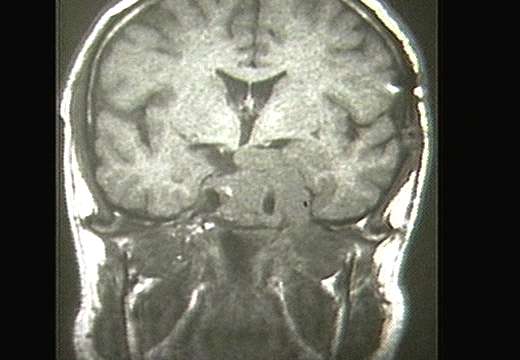
Prolactinoma on MRI - Coronal Plane

Features in the Image
Prolactinomas are the most common hormone secreting tumor of the pituitary gland. This MRI demonstrates a huge prolactinoma with compression of the adjacent cortical tissue. In addition, the tumor entirely encase the left internal carotid artery within the cavernous sinus.
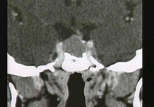
Prolactinoma on CT - Coronal Plane

Features in the Image
This coronal CT shows another prolactinoma considerly smaller than the one shown above on MRI. Even so, it is still growing out of the pituitary fossa and would be considered a macroadenoma because it is bigger than 10mm in diameter. This lesion displaces the infundibulum (aka: pituitary stalk) to the left, but is still largely contained within the pituitary fossa.
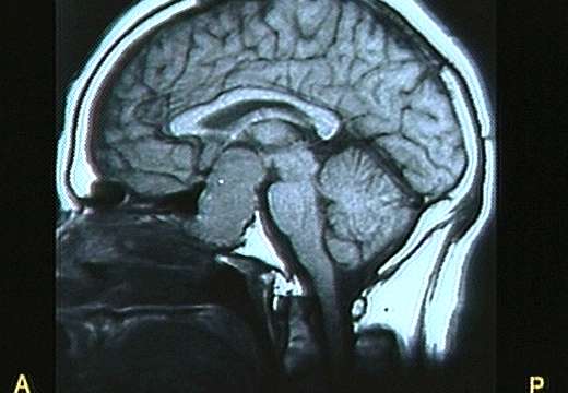
Prolactinoma on MRI - Sagittal Plane

Features in the Image
This large pituitary macroadenoma extends superiorly, anteriorly, and posteriorly out of the pituitary fossa. Anteriorly, it has eroded into the sphenoid sinus. Posteriorly, it has destroyed the dorsum sella, and superiorly, it has diplaced the third ventricle and compressed the optic chiasm.
Forward to Case 16: Renal Mass
Back to Case 14: Microadenoma (Prolactinoma)
Return to Radiology for Medical Students Index
This page hosted by
 Get your own Free Homepage


