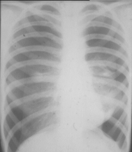
Pneumothorax
The Image

Clinical presentation:
Sudden onset of pleuritic pain and breathlessness in a young adult male. Clinical examination reveals reduced breath sounds, but no dullness to percussion.
Features in Image
There is increased density around the hila with a peripheral well-defined margin. The pulmonary vessels are not seen beyond this margin. The right sided density has a cleft which corresponds to the position of the horizontal fissure. On the left side there is a density marking the site of the lingula.
General features in Pneumothorax
No vessel markings beyond extent of collapse. A white line indicates the lung edge. In tension pneumothorax, there may be marked mediastinal shift. This is a surgical emergency.
Diagnosis
Spontaneous Bilateral Pneumothorax.
It sucks to be tall and skinny. Literally.
Back to Case 9: Neoplasm
Forward to Case 11: Pulmonary Embolus
Return to Radiology for Medical Students Index
This page hosted by
 Get your own Free Homepage
