HOX-Pro DB: THE WAYS OF EVOLUTION OF ENSEMBLES OF HOMEOBOX GENES-CONTROLLERS OF DEVELOPMENT
A.V. SPIROV
The Sechenov Institute of Evolutionary Physiology and Biochemistry, 44 Thorez Ave, St.Petersburg, 194223, Russia; spirov@iephb.ru;
Institute for High-Performance Computing and DataBases, P.O. Box 71, St.Petersburg, 194291, Russia
Nowadays homeobox genes were found for species of main invertebrate and vertebrate phyla. It is established that these genes play key role in time and space orchestration of genome expression during development. Auto- and crossregulatory functional interactions join homeobox genes into genetic networks. For the purpose to orange all available data on structure, functions, phylogeny and evolution of Hox-genes, HOX-clusters and Hox-networks we develop specialized database HOX-Pro.
Its main location is http://www.iephb.ru/~spirov/Hox_pro/Hox-pro00.html. The DB is also mirrored at http://www.mssm.edu/molbio/Hoxpro/new/Hox-pro00.html.
The Hox gene family is highly conserved, yet is responsible for the development of many novel features of the invertebrate and vertebrate body plans. The Hox gene families show progressive expansion by gene duplication in invertebrate species as a tightly linked gene cluster, and further amplification within the chordates primarily by cluster duplication.
Conserved "rainbow-like" expression pattern of the HOX-cluster genes is now well known. However, despite such wonderful functional conservatism, HOX-clusters undergo essential rearrangements in evolution of main phyla. One can distinguish cluster and gene duplications, juxtapositions of duplicated portion of a gene and possible addition of cis-elements by transposons. In our communication we summarize these main ways of HOX-ensembles reorganization as they described in the HOX-Pro. We will stress the interplay between gene duplication and the evolution of gene regulation in the modification of vertebrate body plans.
1. Introduction
Nowadays the Hox genes have become a paradigm for the conservation of developmental mechanisms throughout the animal kingdom. They encode transcription factors that act as molecular markers for the position of cells along the major body axis. Individual Hox genes are activated at different positions in the early embryo, establishing a pattern that is maintained throughout much of development. This differential expression has been shown to control the development of region-specific structures in nematodes, arthropods and chordates, and may be a shared characteristic of triploblastic metazoan animals.
Mutations within homeotic Hox-genes in Drosophila melanogaster transform defined segments of the body into the character of adjacent segments. Homologous genes there are in vertebrates, where it has been possible to mutate them, shifts in morphological character have also been produced.
The discovery of the first HOX cluster outside Drosophila causes great hopes. It was proposed that the homeobox would become a "Rosetta stone" for the study of animal development. This could enable us to read the epigenetic code of other animals on the basis of our understanding of Drosophila [Slack et al., 1993]. It is possible that this HOX-based epigenetic code is very ancient and was in place in the common ancestor of all modern animals. According to Slack, Holland and Graham, this character should be adopted as the defining character, or synapomorphy, of the kingdom Animalia.
We develop the HOX-Pro database for description of homeobox-genes controlling embryogenesis [Spirov, 1996]. It contains a broad spectrum of information including pictures, schemes, and movies. Graphical representation of HOX-clusters and Hox-based networks is accomplished in the form of flow diagrams, as well as in the form of the Java applets, that permits to emphasize the interacting genes in the network, to reflect the mode of gene actions, etc. [Serov et al., 1998].
The distinctive feature of the HOX-Pro is its ability to serve not only as an information depository but also as tool for derivation of new knowledge by means of computer analysis. This communication is intended to show perspectives of comparative evolutionary approach to study functions and evolution of homeobox genes-controllers of development as it is presented in the HOX-Pro.
2. HOX-clusters and Hox-networks evolution
The Hox gene family is highly conserved, yet is responsible for the development of many novel features of the vertebrate body plan. The Hox gene family shows progressive expansion by gene duplication in invertebrate species as a tightly linked gene cluster, and further amplification within the chordates primarily by cluster duplication.
Two explanations have been proposed for the existence of multiple copies of genes. One explanation is that copy number increased by tandem duplication [Forey P & P Janvier. 1994]. This could occur by: (1) unequal crossing over between homologous chromosomes in meiosis; (2) unequal exchange between sister chromatids during mitosis; or (3) regional redundant replication of DNA. In all cases, tandem duplication leads to duplicate linked genes. The second rather extreme explanation is that gene duplications occurred by way of one or more tetraploidization events [Ohno, 1970; Lundin, 1993; Ohno et al., 1986].
According to Susumu Ohno vertebrates arose as a result of two tetraploidization (genome duplication) events [Ohno, 1970]. These events were speculated to have occurred (1) after that first primitive chordates arose (~500 million years) and (2) about the time that the tetrapods (four-limbed vertebrates) arose (~375 million years).
What really happened at the end of the Precambrian or beginning of Cambrian to produce 'known' dazzling array of body plans? The last two decades, a great deal of attention has been focused on Hox genes as a possible explanation for the Cambrian explosion.
The data available permit modern molecular evolutionists to frame several possible scenarios for the relative timing of the evolution of the Hox genes and of the body plans of animal phyla near the time of the Cambrian explosion [Erwin et al., 1997].
It is possible is that lineage divergence, Hox-gene duplications and body-plan formation were spread through the 35-million-year period just before the Cambrian explosion. The last ancestor common to vertebrates and arthropods could have lived nearly 565 million years. Developmental controls in this ancestor presumably evolved first, reaching a level of sophistication that permitted the rise of major morphological innovations and culminating in the explosion of body plans during the late Neoproterozoic and early Cambrian [Erwin et al., 1997].
As a first step towards a developmental history of animal architectures, we can begin to reconstruct the evolution of the Hox clusters using information from developmental/molecular biology and knowledge of the relationships between different phyla.
While overviewing the ways and mechanisms of the Hox genes evolutionary expansion, we must distinguish cluster and gene duplications, juxtapositions of duplicated portion of a gene and possible addition of cis-elements by transposons. Below we summarized these main ways of HOX-clusters and networks reorganization.
2.1. HOX-clusters duplications
The clustered organization of Hox genes provides a powerful opportunity to examine gene gain and loss in evolution because physical linkage is a key diagnostic feature which allows homology to be established unambiguously. Despite suggestions that changes in Hox gene number played a role in evolution of metazoan body plans, there has been a general lack of evidence for such variation amongst vertebrates and it has therefore been widely assumed that differential regulation may be the key element in all vertebrate Hox evolution.
However now situation became drastically change. Although not all phyla have been studied, and the quality of the data remains variable, HOX clusters within phyla that have been well studied are distinctive, produced by unique patterns of gene duplication and loss.
Only one cluster has been found in all insects examined. Only one HOX cluster was initially found in the cephalochordate amphioxus [Pendleton et al., 1993; Holland et al., 1994].
Despite the consensus that a primordial HOX cluster arose by tandem gene duplication close to animal origins, several homeobox genes are found outside the HOX cluster and are known as 'dispersed' Hox-like genes; these genes may have been transposed away from an expanding cluster. Three of these dispersed homeobox genes form a novel gene cluster in the amphioxus [Brooke et al., 1998]. This 'ParaHox' gene cluster is thought to be an ancient paralogue (evolutionary sister) of the HOX gene cluster; the two gene clusters arose by duplication of a ProtoHox gene cluster. But what is more, the amphioxus ParaHox genes have co-linear developmental expression patterns in anterior, middle and posterior tissues. duplication facilitated an increase in body complexity during the Cambrian explosion.
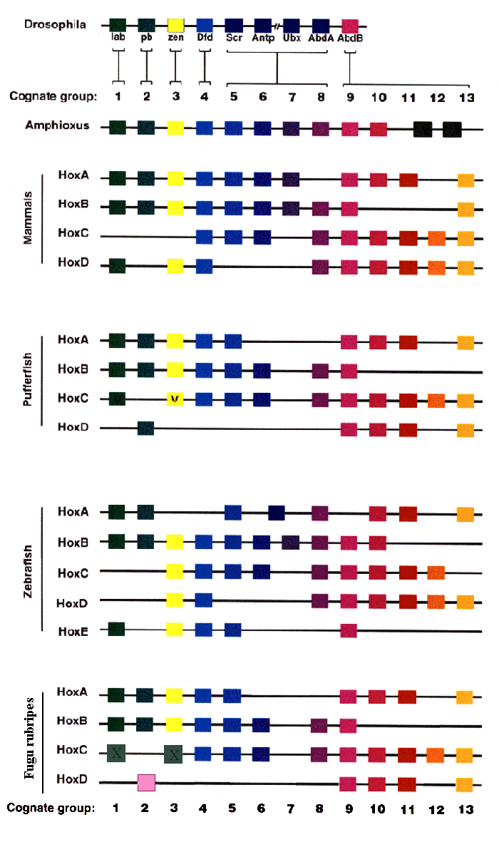
Preliminary findings indicate that the lamprey (Petromyzon) and the horned shark (Heterodontus) are two-cluster species, while the hagfish (Eptatretus) and the ratfish (Hydrolagus) have four clusters. But what is more, zebrafish (Danio rerio) has five (!) HOX clusters (fig.1).

Figure 1. Comparison of insects, cephalohordata, teleost and mammals HOX clusters. The scheme depicts our current knowledge of Hox cluster evolution from invertebrates to mammals.
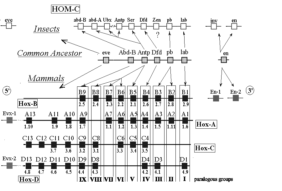
Six Hox genes are shared between mice and flies, indicating that their common ancestor, which lived before the Cambrian explosion, had a HOX cluster composed of at least six genes (fig.2). As living arthropods have eight Hox genes, two of them must have originated by duplication after the divergence of the ancestors of arthropods and vertebrates. Today these two newer Hox genes mediate the development of segments in the middle of the arthropod body. Hox genes mediating the development of the midbody, in addition to other developmental features, were also duplicated within the lineage leading to mammals.
Four HOX clusters in mammals arose presumably by three duplications of one ancestral cluster [Bailey et al., 1997]. By the conserved gene organization in each cluster and criteria of sequence conservation, it is possible to identify closely related groups of genes on different clusters - so called paralogous groups (fig.1). Nine such groups (paralogous groups 1 through 9), are thought to be represented in the ancestral cluster. Nine of assumed 36 genes in these paralogous groups are missing.
To test the possibility that Hox organization may have varied since the origin of jawed vertebrates the Hox gene clusters of a teleost fishes, Fugu rubripes and Fundulus heteroclitus, was studied in comparison with another teleost test-object zebrafish.
Aparicio with co-workers [1997] have identified four HOX complexes in Fugu and found an unprecedented degree of variation when compared with tetrapod clusters. Fugu clusters are widely variant with respect to length; at least nine genes have been lost; there is a new group-2 paralogue; and pseudo-gene remnants of group-1 and group-3 paralogues were found in the HOX C complex, when compared with the present mammalian clusters (fig.1). The putative Fugu HOX D complex illustrates the most extreme example of all the HOX clusters, when compared with mammalian clusters: five genes are missing and the 5' linkageis with hydroxy butyrate dehydrogenase enzyme rather than Evx2. It is possible that some fish had, and may still have (as zebrafish), more then four HOX complexes and that Fugu has lost the cognate HOX D counterpart [Aparicio et al., 1997].
In the teleost fish Fundulus heteroclitus, a total of 22 members from eight paralogous groups were found, consistent with as many as ten of 32 genes missing [Misof and Wagner, 1996]. The number of representatives in cognate groups 1, 4, 5, 6, 7, 8 and 9 differed from those of human and mouse (fig.1). For groups 1 and 9 four representatives were found. It demonstrates significant differences of the Fundulus clusters compared to those of mouse and humans.
In the teleost zebrafish there are five HOX clusters with as many as ten missing genes in paralogous groups 1 through 9 [Misof et al., 1996]. Prince with co-workers has isolated 32 zebrafish Hox genes. In comparison to the tetrapods, zebrafish has several additional Hox genes, both within and beyond the expected 4 HOX clusters (A-D). For example, a member of Hox paralogue group 8 lying on the HAX A cluster, and a member of Hox paralogue group 10 lying on the HOX B cluster, have no equivalent mouse or human genes (fig.1). Beyond the 4 clusters (A-D), a further 3 Hox genes (the Hoxx and y genes) was isolated, which lie in paralogue groups 4, 6, and 9. The Hoxx4 and Hoxx9 genes occur on the same set of hybrid chromosomes, hinting at the possibility of an additional HOX cluster for the zebrafish.
These findings show that gene loss after duplication of the prototypical vertebrate HOX clusters is a key feature of both tetrapod and fish evolution. Comparison of HOX clusters organisation for known fish and tetrapod Hox ensembles gives good illustration of non-random lack of paralogous genes in the HOX clusters, A - D (Table 1). According up-to-date knowledge, the most conservative in tetrapod and fish evolution are assumed to be HOX B and HOX C clusters (Table 1). Tetrapods and teleosts all have eight HOX B genes, from the first to ninth paralog, and eight HOX C genes, from the fourth to twelfth paralog. In both these conservative genomic sequences the seventh paralogous gene can be lost. While HOX A and HOX D clusters are essentially more variable. In particular, all up-to-date investigated vertebrates lost three HOX D genes from fifth to eighth paralogs.
Table 1. Pattern of lacking of paralogous genes in four HOX clusters of four vertebrate species
| Cognate
groups |
I | II | III | IV | V | VI | VII | VIII | IX | X | XI | XII | XIII |
| HOX-A | - | - | 1 | 1 | - | 3- | 3- | 3 | 1 | - | - | 4 | - |
| HOX-B | - | - | - | - | - | - | 2 | - | - | 3 | 4 | 4 | 3 |
| HOX-C | 2' | 4 | 1' | - | - | - | 4 | - | - | - | - | - | 1 |
| HOX-D | 3 | 2 | 2 | 2 | 4 | 4 | 4 | 2 | - | - | - | 2 | - |
The number inside each cell indicates the number of cases of lost genes for given cluster and given paralogous group.
Apart from physical missing of the Hox genes we must mention the cases of functional lost of the genes. Within the HOX-cluster of Arthropods homeotic genes the two Anip-like genes which lack the function of the genes-selectors could be pointed out. These genes are fushi tarazu (ftz) and zerknult (fig.1). zen genes of Drosophila derive from a Hox class 3 sequence that formed part of the common ancestral Hox cluster, but in insects this Hox gene has lost its role in patterning the anterio-posterior axis of the embryo, and acquired a new function. In the lineage leading to Drosophila [Falciani et al., 1996], the zen genes have diverged particularly rapidly, as opposed to the other genes from the same cluster. The fundamental role in the processes of divergention of the main groups of arthropods is also ascribed to them.
In connection with this, another rapidly evolving gene, even-skipped (eve) should be mentioned. Homeodomain of this gene demonstrates the homology with the same of ftz and Antp. This gene controls the process of neurogenesis in arthropods. Beyond this, supposedly, ancient function of this gene, eve appears to be involved in the processes of segmentation of the early embryo of the Diptera. However, eve gene of Diptera is not linked with the HOX-cluster, whereas its homologues of vertebrates are localized in the HOX A and HOX D clusters (fig.1) as the 5' end genes. Possibly, during the evolution in the ancestors of arthropod eve lacked the physical and functional relations with the homeotic cluster, while at the evolution of vertebrates, such event has not take place. That is why, the rapidly evolving genes of the HOX/HOM-cluster are of particular interest from the positions of the molecular evolution.
One more example, the Hox gene engrailed is important in controlling segmentation in chordates, arthropods, and annelids, but in annelids it controls nervous system segmentation (see Patel et al., 1989), while in arthropods and chordates it controls body segmentation (but with different roles; see Osorio et al., 1995).

Figure 2. Possible evolutionary relationships between insect and vertebrate clusters of homeobox genes. Alignment of the four vertebrate HOX clusters with the Drosophila HOM-C homeotic complex is presented. The letters above the boxes represent modern nomenclature. Below the boxes are the former mouse gene names.
2.2. Juxtaposition duplicated portion of a gene to new cis-regulatory elements
According to Li and Noll, during duplication duplicated portion of a gene, including its coding region, is juxtaposed to new cis-regulatory elements which change its expression and thus give it a new function - ultimately the activation of different set of genes [Li and Noll, 1994].
The three paired-box and homeobox genes paired, gooseberry and gooseberry neuro have distinct developmental functions in Drosophila embryogenesis. However, despite the functional difference and the considerably diverged coding sequence of these genes, their proteins have conserved the same function [Li and Noll, 1994]. The finding that the essential difference between genes may reside in their cis-regulatory regions exemplifies an important evolutionary mechanism of how function diversifies after gene duplication.
Other three clustered Drosophila genes homeotic spalt, spalt adjacent, spalt related, and their present cis-regulatory regions arose through a chromosomal rearrangement involving local duplication and transposition events in the 32F/33A region on the left arm of the second chromosome [Reuter et al., 1996].
Conservatism of the translated and untranslated sequences of HOM/HOX-complex genes is well known. However there are impressive exclusions demonstrating evolutionary recent gene reconstructions. In contrast to the coding region, the 5' untranslated region of the mouse Hoxb-3 (Hox-2.7) cDNA differs considerably from that of a human one. Alternative ORF gives transcript encoded predicted protein of 140 amino acids with homology to the ß-chain of mammalian ATPase enzymes (Sham et al., 1992). This finding opens possibility that a transcription unit for a non-homeobox containing gene is located in the HOX B cluster, as observed for transmembrane protein amalgam (Seeger et al., 1988) in the Drosophila ANT-C complex.
2.3 Addition and distribution of control elements by transposons
Transposable elements (TEs) - transposons and retroposons, are a major source of genetic change, including the creation of novel genes, the alteration of gene expression in development, and the genesis of major genomic rearrangements [Lozovskaya et al., 1995].
TE insertions can affect the expression patterns of endogenous genes by adding and distributing specific control elements throughout the host genome. Transposition of a TE into or near a particular host gene - possibly followed by an excision event leaving behind the TE's regulatory sequences - might impose novel developmental control on this host gene. Recent observations have shown that a number of sequences known mobile elements were frequently inserted into the DNA of a gene regions and can influence the regulation of a gene's expression [Britten, 1997].
There are above twenty examples from Drosophila, sea urchin, human and mouse genomes that meet the following criteria [Britten, 1997]:
the element was inserted far in the past and thus the event is not a transient mutation (1);
the element is a member of a large group of similar sequences (2);
the inserted element now serves a useful function (3).
All these cases are summarised in electronic table at http://www.iephb.ru/~spirov/Hox_pro/te-table.html.
The four known Spec genes of sea urchin Strongylocentrotus purpuratus have "RSR" repeated sequences in their 5' regions [Britten, 1997]. The "RSR" element of the Spec2a gene has been examined carefully and includes an enhancer. The RSR enhancer is required for the tissue-specific expression of the Spec2a gene and contains four Otx binding sites for a bicoid-class homeodomain protein of sea urchins (SpOtx).
Two Intracisternal A-Particle (IAP, transposable element, the family of intracisternal A particles) insertions have been identified in WEHI-3B myeloid leukemic cells, one at the interleukin-3 gene and another at the homeobox gene HoxB-8 (Hox-2.4) Aberdam et al,, 1991]. The provirus has inserted within the first exon of the gene and generated a Hox-2.4 mRNA with a 5' sequence derived from the IAP long terminal repeat. Both proviral insertions have resulted in transcriptional activation of the adjacent genes, which appears to be a significant step in the leukemogenic process in these leukemic cells Aberdam et al,, 1991].
On the other hand, Pig Hox B8 gene contains 5' part of L1 repeat [Baay and Miller, 1989]. This Sus scrofa's L1Ss interspersed repeat is related to human LINE1 [http://www.iephb.ru/~spirov/Hox_pro /est_b8-l1ss.html].

Figure 2. Physical map of human HOX A cluster including evx1 gene [Bradshaw et al., 1998; Jones et al., 1998].
There are about sixty of Alu repetitive elements in 3' flanking region (the left side of the picture) and about 20 of Alu repeats in 5' flanking region, between Hox A13 and Evx1 genes (the right side of the picture).
The EST (Expressed Sequence Tag) Project (Washington University) aimed at production of 5' and 3' terminal sequences from normalized cDNA libraries yielded 18 cases of transcripts similar to homeobox protein mRNAs containing Alu and other interspersed repetitive elements (../public_html/new/te-Hox.html). Among these human endogenous retrorvirus HERVFH21I sequence was found in Hox B5 transcripts, while human mosaic endogenous retrovirus HARLEQUIN sequence was found in Hox D4 transcripts.
Vansant and Reynolds [Vansant and Reynolds, 1995] have observed that Alu sequences include functional binding sites for retinoic acid (RA) receptors. The question is risen about the possible function thousands of retinoic acid receptor-binding sites in Alu sequences scattered throughout the human genome. On the other hand, there are well documented examples of mammalian/human genes that include in their promoters Alu sequences carrying the RA receptor- and estrogen receptor-binding sites (Britten, 1997).
To date, the RA-response elements (RAREs) identified in the vertebrate HOX-clusters are RARE of HoxA-1 (murine and human), RAREs of the murine and human HoxB-1 genes and RAREs conserved in the promoters of the human and murine HoxD-4 genes. All these data is summarised at http://www.iephb.ru/~spirov/Hox_pro/Hox-rares.html.
Recently human HOX A cluster with vast 3' flank was completely sequenced (fig.2) [Bradshaw et al., 1998; Jones et al., 1998]. 3' and 5' flanks of the cluster are full of Alu and other repeats (MER, MIR, L1). The 49 kb length intergenic region from gene WUGSC:H_DJ0167F23.5 to Hox A1 contains 56 Alu repeats. It is more than one Alu per one kb! 3' vicinity of the Hox A1 don't include Alu but contains well characterised retinoic acid-responsive enhancer of the cluster [Langston et al., 1997]. This RAIDR5 consists of two direct target sites GGTTCA and AGTTCA with 5-bp spacer.
-GGTTCA-
alu_59091. -GCTGGGCACGGTGGCTCACACCTGTAATCCCAGCACTTTGGGAAGCAGAGGCAGGC
alu_59854. GGTCGGGCACGGTGGCTCATGCCTGTAATCCCAGCACTTTGGGAGGCCAGGGCGGGT
alu_62808. --CTGGGCGCGGTAGCTCACGCCTGTAATCCCAGCACTTTGGGAGGCCGAGGCAGGC
alu_64265. GGCTGGGCACTGTGGCTCACGCCTGTAATCCTAGCACTTTGGGACACCAAGGTGGAT
alu_67146. -GCCGGGGGCAGTGGCTCATGCCTGTAATCCCAGCACTTTGGGAGGCTGAGGTGGGT
alu_69605. GGCTGGGTGCAGTGGCTCATGCCTGTAATCCCAGCACTTTGGGAGGCCAAGGCAGGC
alu_68253. ----GGGCACGGTGGCTCATGCCTATAATCACAGCACTTTGGGAGGCCAAGGCAGGA
alu_69217. GGCCGGGCGCGGTGGTTCACACTTCTAATCCCAGCACTTTGGGAGGCCGAGGCGGGC
*** * ** * *** * * ***** ************ * ** *
-GGTTCA----AGTTCA--AGTTCA-
alu_59091. GGATCACCTGAGGTCGGGAGTTCGAGACCAGCCTGACCAACATGGAGAAACCCCATCTCTAC
alu_59854. GGATCACCTGAGGTCAGGAGGTCGAGACCAGCCTGGCCAACATGGTGAAACCCCATCTCTAC
alu_62808. GGATCA--TGAGGTCAGGAGATCAAGACCGTCCTGGCTAACATGGTGAAACCCCATCTCTAC
alu_64265. GGATCACCTGAGGTCAGGAGTTCAAGACCAGCCTGGCCAACATGGCAAAACCCTCTCTCTAC
alu_67146. GGGTTGCCTGAGATCAGGAGTTCAAGACCATCCTGGCCAACATGGTGAAACCCCATCTCTAC
alu_69605. GGATCACTTGAGGTCAGGAGTTTGAGACCAGCCTGGCCAATATGGTGGAACCCCGTCTCTAC
alu_68253. GGATCACTTGACCTCAGGAGTTCAAGACCAGTTTGGGCAACATGGAGAAACCCTGTCTCTAC
alu_69217. AGATTTCCTGAGGTCAGAAGTTCAAGACCAGCTTGGCCAACATGGTGAATCCTTGTCTCTAC
* * *** ** * ** * ***** ** ** **** * ** *******
Figure 3. CLUSTAL W(1.5) multiple sequence alignment of nine 3' nearest neighbour Alu repeats of human Hox A1 gene. Direct repeats of the target half-sites GGTTCA and AGTTCA are indicated above multiple alignments.
Our sequence analysis show that vast majority of these intergenic Alu repeats belongs to the same group and has conservative sequences containing three direct a/gGTTCA repeats spacered 4(2) and 2 bp (fig.3). On the other side, 19 Alu repeats are scattered at 43-mb 5' flanking region, between Hox A13 and Evx1 genes. Analogously, the most part of these Alu repeats belongs to the same group and has conservative sequences containing three direct a/gGTTCA repeats spacered 4(2) and 2 bp (not shown). All these materials are presented at
This situation is remind the Vansant and Reynolds' results Vansant and Reynolds, 1995]. They show that consensus sequences for the evolutionarily recent Alu subclasses contain three hexamer half sites, related to the consensus AGGTCA, arranged as direct repeats with a spacing of 2 bp, which is consistent with the binding specificities of retinoic acid receptors. These Alu double half sites are shown to function as a retinoic acid response element in experiment Vansant and Reynolds, 1995]. In the case of human HOX A cluster we really have situation with accumulation of Alu repeats containing retinoic acid response elements in the cluster' flanks which likely could alter the expression of the Hox genes.
3. Discussion and Conclusions
Even the cursory inspection of the materials on the functional organization of the Hox-genes and their clusters can lead to some conclusions about mechanisms of their evolution for the main invertebrate and vertebrate groups. The conservatism of the structural and functional organization of the HOX-clusters even during the comparison of very diverged species is well known and has been repeatedly discussed in literature (See [Spirov, 1996]). Hence, the revealed ways and mechanisms of evolutionary variability of the Hox-genes and their ensembles are of fundamental interest.
These overviewed data begin to give us a picture of how body plans, and the genetic machinery that generates them, actually evolved. As multicellular animals emerged, developmental control genes evolved that regulated the architecture of their differentiated bodies. The fundamental purpose of these genes was to mediate the production of various cell, and to array the cell types within tissues and organs in the appropriate order. Doubtless, as body plans became more elaborate and more cell types were required, the gene-regulatory systems were enlarged. The network of genes-controllers apparently was able to alter the pattern of structural gene expressions, rapidly producing distinctive body plans. Thus the genes that direct animal development evolve in the same quirky, opportunistic ways as the morphologies that they produce [Erwin et al., 1977]. Perhaps the relative abruptness with which metazoan body plans were elaborated to produce the Cambrian explosion can be explained by this organizational structure.
Hox genes play a key role in determination of axial and appendicular skeletal morphology and may be a key component of the evolution of diverse metazoan body forms. In this connection, the fact of great attention is that the multiple massive duplications of the large regions of the chromosomes, including the HOX-clusters was accompanied not only by duplication, but also by the essential lack of the Hox-genes.
Particularly, teleost fishes display remarkable variation in skeletal morphology. The diversity of bony forms in the fishes includes the absence of ribs in the order Tetraodonntiformes to which Fugu belongs [Aparacio et al., 1997]. It is plausible that the variation in teleost morphology may be accounted for by variation in Hox gene complement, at least in part.
Above discussed evolutionary conservatism of vertebrate HOX B and C clusters should be compare with the results of phylogenetic trees analysis [Bailey et al., 1997]. Statistical tests of trees of the mammalian HOX cluster genes support ((A,D)(B,C)) un-rooted topology of the tree. Meanwhile the usage the sea urchin and sponge homologue of human DNA sequences as outgroup (og) the rooted tree has the topology (og(A(D(B,C)))), which suggests that the four cluster situation was attained by a three step process. It seems that evolutionary younger clusters B and C remained less diverged than older A and D.
Meanwhile it is the HOX A and D cluster genes that control both teleost fin and tetrapod limb morphogenesis. One could hypothesized that this variability of the A and D clusters is related to divergence in "bauplan" of fish and mammals appendages.
As HOX cluster duplication takes place, some of the rapidly evolving genes can physically and functionally move to the other loci and to the other genetic networks (as it possibly took place in the case of eve and Hoxb-3 paralogous genes in the invertebrate evolution). During the evolution from the lower insects to the higher Diptera, eve gene, supposedly, returned to the segmentation genetic network, but remained outside the HOX-cluster (unlike the two paralogous mammalian genes Evx-1 and Evx-2 which are adjacent to the HOX-clusters).
The evolutionary instability of the HOM-complexes of arthropods and HOX-clusters of vertebrates could be evidently proved by the evolutionary recent insertions (such as the insertion of the fragment of the DNA, encoding the polypeptide similar to ß-subunit of the human ATP-ase into the mouse Hox-2.7 gene Sham et al., 1992]).
It is well known that the Hox-ensembles of vertebrates differ from the same of invertebrates by the key role of the retinoic acid in the control of the Hox-genes expression. On the other hand, the Alu sequences, represented in the vertebrate genomes by many tens thousands of copies, are sometimes carrying the functional RA-dependent enhancers. This fact together with the similarity in sequences of known RA-dependent enhancers in the mammalian Hox-genes with some human Alu sequences, pointed out in the HOX-Pro, leads to assumption about the possible role of the Alu elements in the multiplication and distribution of the RA-responsive elements in the mammalian homeobox-genes.
Acknowledgments
This work is supported by Russian Foundation for Basic Researches (Grant No 96-04-49349).
References
Any comments to aspirov@geocities.com