|
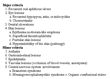
DEFINITION. Behcet's disease is an inflammatory disorder of unknown etiology characterized by recurrent oral and genital aphthous ulcers, ocular inflammation, and skin lesions of erythema nodosum and acneiform eruptions. Behcet's disease also frequently involves the joints, the central nervous system, and the gastrointestinal tract. INCIDENCE AND PREVALENCE. Behcet's disease is common in northern Japan, Korea, China, and the Middle East. In Japan the current prevalence is 1 per 10,000. The frequency in the United States is far less. An annual incidence of 1 per 300,000 was determined for Olmstead County, Minnesota. The disease does not occur frequently in Japanese Americans, perhaps suggesting environmental factors in addition to genetic predisposition in the etiology of this illness. ETIOLOGY AND PATHOGENESIS. No infectious agent has been consistently isolated, although a viral etiology is suspected. Sera frequently contain circulating immune complexes of the IgA and IgG variety, as well as elevated levels of chernotactic activity for leukocytes. Antibodies reactive against oral mucosal cells have been found, and factors produced by patient's lymphocytes are toxic for oral mucosal cells. However, these findings are also present in individuals with recurrent aphthous stornatitis alone. Serum complement levels in Behcet's disease are usually elevated, particularly levels of C9. Most patients with neurologic involvement have demyelinating antibodies in their sera. There is a strong association of HLA-135 with Behqet's disease in Japan and in the Mediterranean area. Heavy metal exposure, certain foods (particularly English walnuts), and toxic factors such as organophosphates have initiated attacks in some individuals. PATHOLOGY. Behqet's disease is primarily an inflammatory disorder involving small blood vessels, particularly venules. Areas of ulceration initially show an intense mononuclear cell infiltration around blood vessels. As the lesion evolves, polymorphonuclear leukocytes and plasma cells predominate. The early lesions resemble a delayed hypersensitivity reaction, the later lesions an immune complex-Arthustype reaction. The role of immune complexes in causing the venulitis is, however, questionable, since immunoglobulins are not routinely found in vessel walls. CLINICAL MANIFESTATIONS. Behcet's disease can occur in many forms, but recurrent oral aphthous ulcers are present in 99 per cent of patients, and in almost 70 per cent of these they are the initial symptoms. Ocular symptoms occur in 90 per cent, skin lesions in 85 per cent, and genital ulcerations in nearly 70 per cent. Arthritis is present in approximately one half of the patients with Behqet's disease. The onset is usually in the third or fourth decade. Behcet's disease affects men and women approximately equally. Indications of poor prognosis include neurologic and posterior uveal tract involvement. In the absence of these, the disease tends to be unpredictable and remitting. In Japan, the mortality is approximately 4 per cent, with blindness occurring in as many as 65 per cent of untreated individuals. Young males have the worst prognosis. TABLE 453-1.
DIAGNOSTIC CRITERIA OF BEHCET'S DISEASE (SYNDROME) The diagnosis of Behcet's disease is based upon the criteria listed in Table 453-1. Complete Behcet's syndrome is associated with all four major criteria. Persons with three major sites of involvement, or ocular lesions plus one other major site, have incomplete Behqet's disease. The diagnosis should be suspected when two major sites are affected. Since all manifestations need not appear, some investigators have grouped this illness into subtypes based upon the primary tissue involvement (i.e., neuro-Behcet's, oculo-Behcet's). The oral aphthous ulcers are painful, unlike those of Reiter's syndrome, and occur singly or multiply on the lingual, gingival, buccal, or labial mucosal membranes. In Reiter's and Stevens-Johnson syndromes, the ulcers occur on the palate, pharynx, and tonsils, structures rarely involved in Behqet's disease. The aphthae of Beh~et's disease usually last for approximately a week and may heal with or without scarring. Ulcers can also appear on the scrotum, vulva, penis, vaginal mucosa, or perianal areas. These lesions may be painless in women. Their gross appearance is similar to that of the oral aphthous ulcers. Vulvar lesions frequently occur premenstrually. Other types of cutaneous involvement are common. Painful, recurrent lesions of erythema nodosum may appear in crops over the tibia. Superficial thrombophlebitis can occur in the upper or lower extremities. Skin eruptions resembling acne vulgaris frequently appear on the upper thorax and face Approximately 40 per cent of patients with Behcet's disease exhibit a cutaneous phenomenon termed "pathergy" and develop sterile pustules at sites of trauma. Venipuncture or injection of sterile saline into the skin of these patients results in the formation of a pustule. This phenomenon is not pathognomonic for Behqet's disease. Ocular lesions may consist of anterior or posterior uveitis. Anterior uveitis frequently produces hazy vision as an initial manifestation. The development of a hypopyon is not unusual. Recurrent posterior uveitis (retinal vasculitis) is an ominous expression of Behcet's disease which, if untreated, frequently leads to bilateral blindness. Choroidal exudates and bleeding may be seen. Articular manifestations consist of arthralgias and arthritis. The involvement is usually asymmetric, affects one to several large joints such as the knees, ankles, elbows, and wrists, and resolves during remissions. Permanent joint damage is rare. Gastrointestinal involvement during acute attacks, present in approximately 50 per cent of patients, is most commonly manifested by vomiting, abdominal pain, diarrhea, flatulence, or constipation. More specific for Behcet's disease are erosions or superficial ulcers in the terminal ileum or colon. Intestinal ulcers occasionally perforate. Differentiation of Behcet's disease from ulcerative colitis or regional enteritis may be difficult. Nervous system involvement occurs in approximately 10 per cent of patients and can be extremely severe, explosive, and associated with a poor prognosis. Manifestations include herniplegia, paraplegia, cerebellar dysfunction, and psychologic changes. Superficial venous occlusions, perhaps related to abnormalities in the blood fibrinolytic system, occur in up to 40 per cent of patients. Inferior or superior vena caval obstructions can lead to death. Occlusive lesions have also occurred in the aorta and other large arteries. Epididymitis occurs in approximately 6 per cent of male patients. TREATMENT. No therapy has been proved uniformly effective. Since the illness is unpredictable and frequently remitting, long-term continuous therapy with potentially dangerous drugs is not justified except in specific situations. Chlorambucil* (0.1 to 0.2 ing per kilogram per day) has been reported to prevent blindness in patients with posterior retinal involvement. Other immunosuppressive agents such as azathioprine,* cyclophosphamide,* and 6-mercaptopurine* have also been used. Cyclosporin* is reported to be effective in abrogating ocular as well as other disease manifestations. Since neuro-Behcet's syndrome is life-threatening, immunosuppressive agents are frequently used for this manifestation. Corticosteroids are strictly palliative for the inflammatory lesions (i.e., anterior uveitis) and should not be used except during acute flares of the disease. Avoidance of factors known to precipitate attacks (i.e., particular foods or toxic materials) should be encouraged. Colchicine* (0.6 mg orally twice a day) is sometimes effective in treating the mucocutaneous and cutaneous lesions. Transfer factor has not been effective in double-blind clinical trials. In patients with gastrointestinal manifestations, a trial of sulfasalazine* (2 to 4 gin per day') is warranted. Sulfasalazine may also be useful in patients without obvious gastrointestinal complaints. Fibrinolytic agents have been recommended for patients with occlusive vascular disease. Because the disease is remitting in nature, the efficacy of therapy is difficult to evaluate. *This use is not listed in the manufacturer's directive |
ENTRANCE | HOME | 1 | 2 | 3 | 4 | LINKS | FUN STUFF | BULLETIN BOARD | BOOK STORE | DISEASES | SEARCH
