BACTERIAL ARTHRITIS
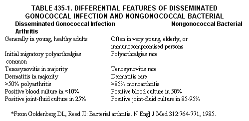
Bacterial arthritis usually results from bloodborne infection and much less commonly from direct penetration (e.g., needle aspiration) or contiguous osteomyelitis. Acute bacterial joint infections may be divided into two general groups, nongonococcal and gonococcal, based on their typically differing target populations, clinical characteristics, and ease of treatment. Differential features of these two classes of bacterial arthritis are presented in Table 435-1.
Nongonococcal Arthritis
Staphylococcus aureus heads the list of common infecting organisms in
this group, followed by other gram-positive cocci (Streptococcus pyogenes,
pneumoniae, viridans) and gramnegative bacilli (Escherichia coli, Salmonella
sp., Pseudomonas, etc.); Haemophilus influenzae is unusual except in children
under four years of age, before protective immunity develops. Patients
are often very young, elderly, immunocompromised, or users of intravenous
drugs. Risk factors for bacterial arthritis during septicemia include
debilitating chronic disease, immunosuppressive therapy, previous joint
damage (e.g., rheumatoid arthritis, neuropathic arthropathy, joint surgery),
sickle cell anemia, hypogammaglobulinemia, and intra-articular corticosteroid
injections. After prosthetic joint replacement, an increasing problem
has been late infection by organisms of low virulence, such as Staphylococcus
epidermidis.
CLINICAL MANIFESTATIONS.
A patient may present typically with the abrupt onset of a single, severely
tender, red-hot swollen joint, especially the knee or another weightbearing
joint; shaking chills and fever may occur. However, signs of inflammation
may be masked in severely debilitated patients or in those receiving adrenocorticosteroids
or immunosuppressive agents. Bacterial arthritis superimposed on a noninfectious
inflammatory joint disease may also be easily overlooked. For example,
infection in one or a few joints of a patient with rheumatoid arthritis
may be mistaken for a flare in the chronic disease. An infected joint
in a gouty individual may go unrecognized for too long, even when the
patient does not respond to his usual regimen for acute gouty arthritis
(a good clue that something else is going on). A high index of suspicion
is essential in these circumstances, because delay can lead rapidly to
destruction of cartilage and bone and eventual fibrous or bony ankylosis.

DIAGNOSIS. When bacterial arthritis is suspected, prompt joint
aspiration and both Gram stain and culture of synovial fluid are imperative;
most nongonococcal bacteria will be recovered. Cultures for both aerobic
and anaerobic organisms should be made. Synovial fluid leukocyte counts
are frequently greater than 50,000 per cubic millimeter, and the glucose
level low and lactate level high compared with those of serum, but these
findings are not specific for infection. Bacteriologic studies should
of course be extended to blood and other material (sputum, urine, etc.)
from which the infection may have disseminated. On x-ray, only soft-tissue
swelling is likely to be seen during the first week, but evidence of loss
of articular cartilage and erosion of bone may appear rather soon thereafter
in untreated patients
MANAGEMENT. Successful management of bacterial arthritis depends primarily on early institution of appropriate antimicrobial therapy and effective drainage of the joint space. The selection of antimicrobial agents and recommendations regarding dose and duration of therapy are discussed in other areas of the text (Part XVIII). Antibiotics are given parenterally, often in high doses, for two to four weeks or more, depending on the clinical situation and the patient's response. They generally attain adequate levels in joint fluid and need not be given intra-articularly; indeed, the latter procedure may induce a chemical synovitis. For drainage, daily (or even more frequent) closed joint aspiration through a large-bore needle is carried out until fluid no longer accumulates. Open surgical drainage usually can be avoided except when the hip or the shoulder is infected (difficult to evacuate completely by needle); when tissue debris or fibrin interferes with closed aspiration; or when loculations, gross joint destruction, or contiguous osteomyelitis is present. The affected joint should be at rest while inflamed, and mobilized to prevent atrophy when signs of acute inflammation have subsided.
Gonococcal Arthritis
Gonococcal infection is discussed in Ch. 306. The associated arthritis
is by far the most common bacterial joint problem in generally healthy,
sexually active teenagers and young adults, especially in urban populations.
Gonorrhea is more likely to disseminate in women. The risk of dissemination
is particularly high during menses and pregnancy, in the post-parturn
period, and in individuals with genetic deficiency in the terminal components
of serum complement (C5, C6, C71 or C8). Additional features that help
to distinguish gonococcal from other bacterial arthritides include a high
frequency of associated tenosynovitis and rash and of multiple joint involvement,
especially in the wrists and hands. Diagnosis is frequently presumptive
because synovial fluid smear and culture-which requires special media-are
often negative, and corroborating cultural evidence from urethra cervix
' throat, rectum, blood, or skin may be lacking. Highly suggestive diagnostically
is a history of fever and migratory polyarthralgias that progress to frank
oligoarticular arthritis and are associated with tenosynovitis and skin
lesions. The latter are either vesiculopustular on an erythernatous base,
often with necrotic centers, or hemorrhagic. Similar lesions are seen
with arthritis caused by the meningococcus. Response to (even oral) antibiotic
therapy (Ch. 306) and drainage is usually dramatic. Resistance to penicillin
of gonococci that disseminate is uncommon.
Tuberculous Arthritis
The general decline in the frequency of pulmonary tuberculosis in the
western world is reflected in the relative rarity of tuberculous bone
and joint disease. Infection usually reaches the joint from hernatogenous
dissemination to bone and direct extension from an osteomyelitic focus.
Formerly, the classic presentation was chronic low back pain in a child
because of involvement of lower thoracic or lumbar vertebrae, leading
to collapse and sharp-angle kyphosis (Pott's disease). Currently, the
typical target is a tuberculin-positive adult, often without evidence
of pulmonary disease, who presents with chronic, insidious pain and swelling,
usually in a single joint, especially the hip, knee, or wrist; this presentation
is often mistaken for monoarticular rheumatoid arthritis. Tenosynovitis
is common. Diagnosis depends upon culture of Mycobacterium tuberculosis
from synovial fluid (positive in 80 per cent) or synovial biopsy (positive
in 90 per cent). Sensitivities to chemotherapeutic agents must be determined;
caseating granulomata, and acid-fast bacilli are sometimes due to atypical
mycobacteria resistant to the usual antituberculous drugs. Usual therapy
for uncomplicated infections consists of long-term isoniazid and ethambutol
or rifampin. Goldenberg DL, Reed JI: Bacterial arthritis. N Engl J Med
312:764, 1985. A compact, well-referenced review of the pathophysiology
of bacterial arthritis, clinical and microbiologic characteristics of
its common forms, and current approaches to diagnosis and therapy.
VIRAL ARTHRITIS
Many specific viral infections
are associated with polyarthritis, notably hepatitis B and rubella, but
also mumps and vaccinia (and formerly, smallpox) and occasionally adenovirus
type 7, Ebstein-Barr virus (EBV) (in infectious mononucleosis) and other
herpes viruses, and certain enteroviruses. Polyarthritis may dominate
the picture of various mosquito-transmitted arbovirus infections, especially
epidemic polyarthritis of Australia (Ross River virus) and the dengue-like
illnesses, chikungunya and o'nyongnyong. In general, diagnosis is suggested
by the exposure history (drug abuse for hepatitis B, immunization for
rubella, epidemiologic considerations for arbo- or enteroviruses); recognition
of the associated viral syndrome, which often includes fever, rash, and
regional lymphadenopathy; brevity of the joint involvement (days to weeks);
and changing antibody titers against specific antigens.
Routine laboratory tests are nonspecific, and except
for rubella, virus has rarely been recovered from synovial fluid. Little
is known about pathogenesis, but studies of hepatitis B and rubella provide
some clues. Transient, often symmetric polyarthritis or arthralgias resembling
acute rheumatoid arthritis may be associated with both hepatitis B and
rubella infections. In the case of hepatitis B, 10 to 30 per cent of patients
have arthritis, often accompanied by urticaria, fever, and lymphadenopathy,
all occurring days to weeks before the onset of frank hepatitis. This
prodromal syndrome typically occurs when hepatitis B surface antigen (HBsAg)
is in excess over antibody, hypocomplementemia is present, and serum contains
immune complexes composed of HBsAg and anti-HB, other immunoglobulins,
and complement components. Similar material has been ound in affected
dermal blood vessels, and the antigen has been seen in synovial tissue.
With the development of antibody excess, complexes disappear, the arthritis
and rash resolve, and frank hepatitis may supervene. The process resembles
experimental serum sickness and suggests an inflammatory pathogenetic
mechanism driven by deposition of immune complexes. Joint symptoms may
respond dramatically to salicylates.
Rubella arthritis is primarily a disease of adult women.
It usually follows onset of the characteristic rash by a few days, but
the rash may be absent and rheumatoid factor present, inviting diagnostic
confusion. Arthritis is usually sudden in onset, symmetric and polyarticular
in distribution (fingers, knees, wrists), brief in duration (less than
a month), and without residua. Salicylates are useful for pain and stiffness.
Arthritis may also occur within a few weeks of vaccination by attenuated
rubella virus. Again, attacks are brief but may recur periodically for
a few years without permanent joint damage. Rubella virus has been recovered
from synovial fluid in both the natural and vaccine-induced disease and
more recently in a few patients with various chronic joint syndromes.
It seems capable of replicating in synovium; whether its new association
with chronic disease is critical or coincidental remains to be determined.
Steere AC: Viral arthritis. In McCarty DJ (ed.): Arthritis and Allied
Conditions. Ilth ed. Philadelphia, Lea & Febiger (in press). Survey of
common and uncommon arthritides associated with specific viral illnesses.
Wands JR, Mann E, Alpert E, et al.: The pathogenesis of arthritis associated
with acute hepatitis B surface antigen-positive hepatitis. Complement
activation and characterization of circulating immune complexes. I Clin
Invest 55:930, 1975. Clinical description and characterization of immunologic
aspects of the syndrome.
OTHER FORMS OF INFECTIOUS ARTHRITIS
Lyme Disease (See Ch. 313.)
Syphilitic Arthritis
Syphilis is discussed in Ch. 310. joint disease associated with congenital
and acquired syphilitic infections is now rare. In infants with congenital
disease, musculoskeletal complaints are related to periostitis and osteochondritis.
About the time of puberty, painless knee effusions (Clutton's joints)
may be confused with rheumatoid or pyogenic arthritis. With acquired infection,
arthralgias, arthritis, or tenosynovitis may accompany classic signs of
secondary syphilis: rash, mucous plaques, alopecia, or lymphadenopathy.
In tertiary lues, gummatous arthritis or periostitis (tibia, clavicles)
may occur. Neuropathic arthropathy (Charcot's joint) is reviewed in Ch.
482.
Fungal Arthritis
Any of the invasive mycoses can affect joints, usually by direct extension
from bone. Frequent infectious agents include coccidioidomycosis and histoplasmosis-both
of which may also be accompanied by erythema nodosum with joint involvement,
sporotrichosis (often by direct penetration: rose thorns), blastomycosis,
actinomycosis, and candidiasis. Clinically, the affected joint or joints
resemble those in other forms of granulomatous arthritis (e.g., tuberculous).
For diagnosis, the causative agent must be seen in appropriately stained
synovial biopsy material or grown from synovial tissue or fluid.