Systemic lupus erythernatosus (SLE) is a disease of unknown etiology characterized by inflammation in many different organ systems associated with the production of antibodies reactive with nuclear, cytoplasmic, and cell membrane antigens. Individual patients may have some, but not necessarily all, of the following: fatigue, anemia, fever, rashes, sun sensitivity, alopecia, arthritis, pericarditis, pleurisy, vasculitis, nephritis, and central nervous system disease. The course is often unpredictable with variable periods of exacerbations and remissions. There is no one clinical abnormality that definitely establishes the diagnosis, nor is there a single test for the disorder. As a result, criteria have been developed and modified in an attempt to include patients with SLE and to exclude pati nts with other disorders (Table 436-1). Although these criteria were developed for epiderniologic and research purposes, they are helpful in diagnosis as well. Nevertheless, it is possible to fulfill these criteria and not have SLE, and it is possible to fail to fulfill the criteria and still have SLE. Thus, a teen-age girl with a "butterfly" rash of the face, pleurisy, and large amounts of serum antibodies reactive with native DNA undoubtedly has SLE even if she does not yet manifest any other criteria. The disease derives its name (lupus =wolf) from the facial rash, which resembles the malar erytherna of a wolf.
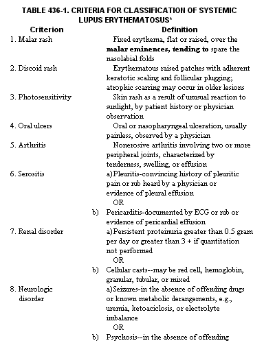
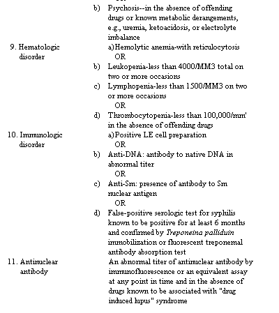
*The classification is based on 11 criteria. For the purpose of identifying patients in clinical studies, a person shall be said to have systemic lupus erythematosus if any 4 or more of the 11 criteria are present, serially or
INCIDENCE. Although SLE can occur at any age (it has been diagnosed at birth and in individuals in the tenth decade of life), more than 60 per cent of patients experience the onset of disease between ages 13 and 40 years. Among children, SLE occurs three times more commonly in females than in males. In patients in their teens, twenties, and thirties, 90 to 95 per cent are female. Thereafter, the female predominance again falls to that observed before puberty. The disorder is approximately three times more common among American blacks than American caucasians. Certain North American Indian tribes (Sioux, Crow, Arapahoe) have an even greater predisposition toward SLE. Orientals have been less well studied; however, the data suggest that they are affected to approximately the same extent as American blacks. The overall annual incidence of SLE is about 6 new cases per 100,000 population per year for relatively low-risk populations and approximately 35 per 100,000 for relatively high-risk populations. The chance of a black female developing SLE in her lifetime is approximately 1 in 250. These data suggest that both genetic factors and sex hormones may affect the probability of developing SLE. If a family member has SLE, the likelihood of SLE increases (approximately 30 per cent for identical twins and 5 per cent for other first-degree relatives). Although males develop SLE less frequently than do females, their illness is not milder.
ETIOLOGY. The etiology of SLE is unknown. The signs and symptoms are thought to be caused by the autoantibodies that react with self constituents and initiate inflammatory responses. The initiation of this process may be multifactorial (Fig. 436-1) and may be different in different individuals. These factors are currently poorly understood. One or more genetic factors appear to be important in many individuals. These may be genes that allow augmented antibody responses following a variety of stimuli as well as genes that predispose to particular autoantibodies. In addition, hormonal, metabolic, and environmental factors appear to act on the genetically conditioned immune substratum to predispose to or protect against disease expression. Males are protected against SLE by their androgens except in a subgroup of males who inherit a Y chromosome accelerating factor from their fathers. Estrogens probably predispose to SLE to a lesser extent than androgens protect against the expression of the illness. In general, factors that augment immunity favor disease expression, whereas those that retard immunity, especially antibody production, tend to protect. Any of a variety of bacterial, viral, or parasitic infections may stimulate the immune system, as may drugs or food additives. Some patients may have a primary abnormality in the ability of their immune systems to perform normal self-regulatory functions. It is probably best to consider abnormal immune regulation as one of several factors that may contribute to illness. If any one is very abnormal, disease may occur. Under most circumstances, several defects probably combine to incite disease. However, once the process is initiated, impaired selfregulation would favor perpetuation of the disease-inducing abnormalities. It has long been known that some individuals with SLE have disease exacerbations following exposure to ultraviolet light. Several different mechanisms are likely: UV light induces keratinocytes to secrete interleukin 1, which, in turn, stimulates B cells and induces T cells to produce growth factors (interleukin 2, B cell growth factors, B cell differentiation factors, interferon), which stimulate the immune system; UV light impairs processing of antigen and immune complexes, thereby increasing the load of pathogenic complexes on target organs; UV light induces cytosine and thymine dimer formation, which stimulates immune responses.
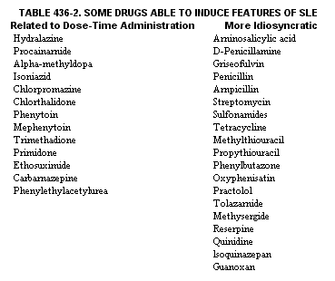
Certain drugs can cause an SLE-like illness in apparently healthy individuals. The drugs (Table 436-2) do not share common structural or chemical properties. The mechanisms of disease induction probably vary. Even chemicals in foods may induce SLE. For example, alfalfa sprouts contain Lcanavanine, which can induce an SLE-like illness. The extent to which "idiopathic" SLE is triggered by such specific environmental factors is unknown. A variety of complement deficiencies have been associated with SLE. The most common is C2 deficiency. It is not clear whether the association is one of genetic linkage or predisposition because of the deficiency itself. The latter might occur if the deficiency led to increased susceptibility to infections that trigger illness. For many years it has been thought that there might be a "lupus virus," a particular virus that induces disease. Patients with SLE have, in the endothelial cells of their kidneys and in their lymphocytes, structures that resemble viral nucleocapsids. In addition, retroviruses have been implicated in the immune-complex renal disease of animals with SLE-like disorders. Nevertheless, even if such a virus is important, it is only one of many factors critical to the development of disease.
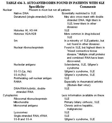
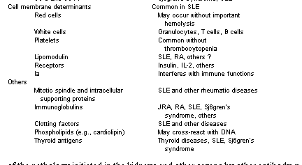
AUTOIMMUNITY AND DISEASE IN SLE. In past years it was believed that an anti-self response was harmful and that such responses did not occur normally. We now appreciate that normal immune responses involve self-self recognition. Moreover, many individuals produce nonpathogenic antibodies reactive with self antigens. As a result, disease occurs only when anti-self reactions are either excessive or productive of especially injurious immune responses. SLE is characterized by the production of large amounts of antibodies reactive with antigens having a great variety of specificities. Some antibody molecules cross-react with more than one antigen (DNA and cardiolipin or IgG and nucleoprotein). As a result, the true range of antibody molecules reactive with self-determinants may be less than the number of specificities as measured by the reactive antigens. Nevertheless, the range of determinants against which SLE antibodies may react is impressive (Table 436-3).
How could such self reactivity come about? It is believed that self-tolerance is a complex state brought about and maintained by several mechanisms. Very early in life, expo sure to antigens tends to produce tolerance rather than immunity. Subsequently, several immune mechanisms maintain self-tolerance. Although B lymphocytes and their progeny are responsible for antibody production, under most circumstances they require helper T lymphocytes for activation, proliferation, and differentiation into antibody-secreting cells. Moreover, specialized T cells (suppressor cells) appear capable of down-regulating immune responses. If the T cell population is self-tolerant, it may prevent B cells from proliferating and differentiating into autoantibody producing cells. A defect in self-tolerance mechanisms could occur at any of several steps in the immune pathway. In addition, strong immune stimulation can overwhelm normal regulatory mechanisms, rendering them incapable of regulating the immune stimuli. Strong immune stimuli such as graft-versus-host disease (as after allogeneic bone marrow transplantation) or stimulation by any of a variety of powerful polyclonal immune activators (endotoxin) or even viruses that stimulate B cells (EbsteinBarr virus) may drive B cells to produce antibodies and autoantibodies without the usual requirements for or regulation by T cells. Individuals with B cells capable of producing pathogenic autoantibodies that had been previously held in check by T cells may, under such circumstances, be driven to produce large amounts of injurious antibodies. Since it is often the quantity of autoantibody that determines whether or not disease occurs, quantitative aspects of immune regulation and immune stimulation may be critical to the balance between disease and relative health with minor immune abnormalities.
PATHOGENESIS. Systemic lupus is often classified as an immune complex type disorder. This designation is, at best, an oversimplification. SLE is a disease primarily mediated by antibodies; however, the details of pathogenesis are not proven for many of the clinical and pathologic findings. It is clear that patients with SLE produce autoantibodies and that many of these are injurious. This has been well demonstrated for the renal disease associated with SLE. Antibody reacts with antigen either in the circulation or in the glomerulus, and complement is fixed, leading to release of chemotactic factors, attraction of leukocytes, and release of their injurious mediators of inflammation. The degree of pathology is determined, to a large extent, by the magnitude of the antibody deposition and the intensity of the inflammatory process initiated. Continued deposition of antibody and continued induction of inflammation ultimately leads to irreversible renal damage. Similar processes occur in other organs.
However, antibody and complement may be deposited in the skin or in the choroid plexus with or without an attendant inflammatory response. The qualitative character of the antibody molecules (affinity, isotype, charge), the nature of the antigen or their combined properties (size, molecular configuration), or additional factors may be critical to pathogenesis. It has long been taught that immunopathology of SLE results from the deposition of DNA-anti-DNA immune complexes. Although immune complexes may contribute to the immunopathology, it is likely that uncomplexed antibody may reach an organ where antigen is already present and there bind to antigen and initiate the inflammatory process. This idea is supported by demonstrations of DNA binding to basement membranes without antibody. Moreover, antibodies of other specificities appear to contribute to disease. Antibody plays a role in SLE not only by depositing in vessels, but also by binding to the surfaces of cells. Patients with SLE produce antibodies to erythrocytes, granulocytes, lymphocytes, and macrophages. These antibodies can cause such cells to be removed from the circulation by the reticuloendothelial system or to be killed by complement-mediated cytotoxicity, or more likely, by the mechanism of antibodydependent cellular cytotoxicity (ADCC). In this non-complement-mediated killing, leukocytes recognize antibody-coated target cells and kill them. ADCC may be responsible for some of the pathology initiated in the kidneys and other organs by other antibody-mediated mechanisms. In addition, antibody directed against renal antigens, for example renal tubular or glomerular basement membrane, may be generated as a result of immunization by fragments released from the inflammatory process. Such antibody induces additional renal pathology and may account for much of the disease in some patients.
It appears that many of the inflammatory lesions that occur in SLE are initiated by antibody and that the injury occurs in small vessels. Thus, any organ so affected could be a site of inflammation with the possibility of scarring, dysfunction, or both. Many of the central nervous system problems of patients (seizures, psychoses) as well as hematologic (anemia, thrombocytopenia, leukopenia), cardiac (coronary artery disease), dermal (alopecia, sun sensitivity), and other clinical and laboratory abnormalities have additional pathogenetic mechanisms. The hematologic abnormalities could all be explained by antibodies specifically reactive with the formed elements of the blood; however, many relate to suppression at the level of the bone marrow. The central nervous system disorders are multiple, and each may have its own pathogenetic mechanism. Because central nervous system involvement in SLE is not a single entity, individual patients may require different approaches to understanding and therapy.
Individual organs often have their peculiar abnormalities. The spleen has "onion skin lesions," concentric fibrosis of the walls and surrounding tissues of the central and penicilliary arteries. These lesions are thought to be diagnostic of SLE in patients with "idiopathic" thrombocytopenia. Nonbacterial verrucous endocarditis (Libman-Sacks) consists of vegetations on the heart valves or chordae tendineae; they can extend along the endocardium and become quite large. The renal pathology varies from mild to severe glomerular inflammation and variable interstitial involvement. Most patients have relatively normal kidneys or a renal lesion consisting of minimal focal hypercellularity, thickening of the capillary basement membrane, and fibrinoid change. In clinically important glomerulonephritis, these lesions are more generalized and are usually a mixture of proliferative and membranous changes with increases in endothelial, mesangial, epithelial, and inflammatory cells, capsular inflammation leading to crescent formation, and focal thickening of the basement membrane and mesangial hypercellularity. Some kidneys have membranous glomerulonephritis with considerable thickening of the basement membrane. Basement membrane thickening, when associated with fibrinoid changes, results in the so-called wire loop lesions. There may also be hyaline thrombi in glomeruli, focal necrosis, hernatoxylin bodies, and sclerosis in healed lesions. Tubular degenerative changes and mixed inflammatory interstitial inflammation are common. Some patients have primarily mesangial disease; this carries a better prognosis than does capillary loop involvement. Extensive crescent formation and substantial glomerular or interstitial scarring are unfavorable prognostic signs.
CLINICAL MANIFESTATIONS
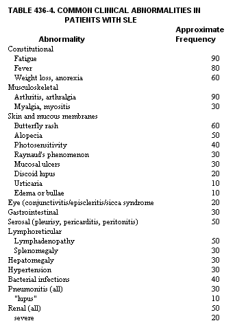
SLE is a highly variable disease in onset and course. A young woman may present with a butterfly rash, a history of recent sun sensitivity, pleuropericarditis, arthritis, fever, extreme fatigue, seizures, and nephrotic syndrome. This "typical" presentation, which is easily recognized as SLE, occurs in only a minority of patients. More commonly, patients may have only one or two signs or symptoms of SLE, such as arthritis and fatigue. Only later do additional features of SLE occur. As a result, the initial presentation may not allow a definitive diagnosis nor insight into the organ systems that may become involved in the future. Many patients never develop major organ involvement. Some have kidney but not central nervous system involvement or vice versa. Thus, the clinical manifestations of one patient may be very different from those of another. Some associations between serologic findings and clinical features are valid on a statistical basis but may not hold for a given individual. Thus, patients with large amounts of antiDNA, especially precipitating antibodies, are more likely to have renal disease. Those with antibodies to Ro (SS-A) and La (SS-B, Ha) are most likely to have sicca syndrome, muscle disease, lung disease, and inconsequential or no renal disease. In the paragraphs that follow, individual clinical features of patients with SLE are described (see also Table 436-4). Patients vary greatly in organ system involvement and also in the severity of disease when a given organ system is affected. Thus, most patients do not have many of the abnormalities described. In addition, SLE is characterized by periods of active disease followed by periods of less intense disease or even remission. In rare cases the patient has a rapidly progressive disease, but the majority can look forward to the time when the disease no longer interferes with their lives.
Constitutional Problems. The majority of patients have fatigue, fever, and weight loss at the time of diagnosis. However, before attributing these to SLE, a diligent search is necessary to rule out infection. Later in the illness, the recurrence of one or more of these findings often indicates an increase in disease activity. Fatigue, difficult as it may be to evaluate, often is the first sign that a flare is imminent.
Musculoskeletal Problems. Arthralgias are the single most common manifestation in SLE. They characteristically are much more transitory than in patients with rheumatoid arthritis (RA), lasting minutes to days in a given joint. With more long-standing or more severe disease, the pain may be constant and frank arthritis is observed. It is often symmetric, the proximal interphalangeal joints of the hands, metacarpophalangeal joints, wrists, and knees being most commonly affected. Morning stiffness is reported by many patients with SLE and joint disease. Although the bony erosions characteristic of RA do not occur, deformities similar to those in RA develop in 10 to 15 per cent of patients and are thought to result from tendon disease. Occasionally patients experience rupture of the Achilles or quadriceps tendons. Myalgias occur in approximately 30 per cent of patients; only a portion of these have muscle tenderness. Many patients with SLE and muscle disease do not have elevations of creatine kinase activity; some of these patients have an elevated aldolase value. Muscle samples from such patients may be normal or show perivascular infiltration or atrophy. Some patients have a vacuolar myopathy that is also observed in corticosteroidtreated individuals.
Skin and Mucous Membranes. The typical butterfly rash varies from a slight blush to a clear-cut and somewhat edematous nonpapular erythernatous covering of both cheeks and the bridge of the nose. Patients with a butterfly rash often look as though they have applied too much rouge. This lesion may occur in the absence of sun exposure, but is often exacerbated by the sun. It often precedes other manifestations of disease. A maculopapular erythematous eruption is also common. Indistinguishable from a drug eruption, it is often induced or exacerbated by sunlight and sometimes by a drug (Gantrisin and ampicillin are common offenders). The palms and soles are not always spared. Healing usually occurs without scarring. Urticaria and angioedema are more common than subepidermal bullae, which occur in only a few per cent of patients. Discoid lupus in SLE is indistinguishable from discoid lupus without systemic involvement; however, systemic disease may develop in patients with long-standing discoid lesions. In this rash, central atrophy, hyper- and hypopigmentation, telangiectasia, and follicular plugging accompany the usual stages of erythema followed by hyperkeratosis and then by atrophy. The hypopigmentation may be extensive and particularly disturbing to blacks. Livedo reticularis occurs commonly in patients with SLE, but only rarely is it severe. Splinter hemorrhages, tender fingertip pulp lesions, and palmar erythema are sometimes remarkable. Purpura is more often secondary to vasculitis or capillary fragility (corticosteroid therapy is often responsible) than to thrombocytopenia. Lupus profundus (relapsing nodular nonsuppurative pan-niculitis) occurs rarely in SLE patients. It may be limited to superficial panniculitis or may extend deeply into the thighs or buttocks. The overlying skin may ulcerate, and the deeper lesions often calcify. One fifth of patients demonstrate vasculitic lesions of the skin. These can occur on the fingertips, forearms, lips, or lower leg (the latter may ulcerate). Although they are signs of disease activity, they do not usually imply impending disaster. The related mucosal ulcers are often painless and occur on the hard and soft palate, the nasal septum, other parts of the upper respiratory tract, and even the vagina. They are usually harmless, but occasional patients with involvement of the upper airway may require emergency tracheotomy. Alopecia is usually diffuse; patients report increased hair on comb or brush or pillow. The hair will regrow in areas not scarred by discoid lesions. Raynaud's phenomenon may be severe enough to cause digital gangrene and spontaneous amputation of the distal parts of the digits. More often it follows a more benign and variable course. Thrombophlebitis occurs in approximately 10 per cent of patients and may be accompanied by pulmonary emboli.
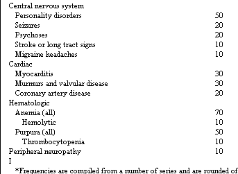
*Frequencies are compiled from a number of series and are
rounded off to the nearest 10 per cent. There was some variation from
series to series depending upon patient population, non-SLE therapy, and
therapy for SLE. Some abnormalities are more common in younger patients
than in older patients (e.g., splenomegaly and lymphadenopathy) and vice
versa (e.g., muscle disease and sicca syndrome).
Eyes. Conjunctivitis or episcleritis or both are usually observed in younger patients at times of disease activity. Cytoid bodies (white exudates next to retinal vessels) are associated with active central nervous system involvement. Spasm of the retinal vessels may lead to transient or permanent blindness. Keratoconjunctivitis sicca occurs in 10 per cent of patients and is usually slowly progressive, but it often improves temporarily with therapy for other symptoms.
Gastrointestinal System. Anorexia, nausea, vomiting, and abdominal pain are observed in a minority of patients. Diffuse abdominal pain with or without rebound tenderness may be a manifestation of serositis or mesenteric arteritis. The latter can be complicated by intestinal infarct, which may lead to perforation and death. Corticosteroid therapy often improves symptoms in both situations; however, if perforation has already occurred, the symptoms may be blunted and therapy inappropriately delayed. Pancreatitis is occasionally due to SLE. Dysphagia may be associated with reduced peristalsis, ulcerations of the esophagus caused by arteritis, or, more commonly, Candida albicans infection.
Liver. Liver enlargement is common but usually inconsequential. Fatty infiltration may rarely be associated with hepatic insufficiency. Liver enzyme elevations often occur early in the illness in the absence of therapy. Aspirin treatment may induce such enzyme elevations. Chronic hepatitis is only occasionally observed in patients with SLE.
Heart. Pericarditis is usually symptomatic but without consequence; however, an occasional patient may experience tamponade. The most common EKG abnormality is nonspecific T wave changes. A prolonged P-R interval or evidence of ischemia or infarction may be found. Myocarditis may be manifested by unexplained tachycardia or mild dyspnea on exertion. More severe involvement is associated with frank heart failure. Coronary artery disease, most often atherosclerotic but occasionally arteritic, can lead to myocardial infarction, even in women in their early twenties.
Lung. Pleuritic chest pain occurs more commonly than does x-ray evidence of effusion; however, massive effusions may occur. Pneumonitis in patients with SLE is often infectious; however a noninfectious syndrome occurs in SLE patients which varies from fleeting infiltrates (usually hemorrhagic) to marked consolidation and hypoxia. Diffuse interstitial pneumonitis also has been found in SLE.
Hematologic and Lymphoreticular Problems. Lymphadenopathy and splenomegaly may be sufficiently marked to suggest a lymphoproliferative disorder. Moreover, polyclonal immune hyperactivity in the lymph node may be mistaken for giant follicular or other lymphomas. Hematologic abnormalities are almost invariably present in patients with active disease. The most common is a normocytic anemia caused by impaired erythropoiesis. Hemolysis may occur in patients with or without a positive Coombs' test result, but significant hemolysis occurs in less than 10 per cent of patients. Iron deficiency often contributes to anemia. Many patients with lupus bruise easily; therapy and capillary fragility are more often the cause than a bleeding disorder. Mild thrombocytopenia occurs in a substantial proportion of patients; however, serious thrombocytopenia occurs in less than 10 per cent. Two types of "anticoagulants" occur. One is a laboratory finding caused by antibodies reactive with the phospholipids used in the partial thromboplastin time (PTT) test. This abnormality is not associated with prolonged bleeding and is not a cause of concern with regard to surgery or biopsies. Other antibodies may react with clotting factors (VIII, IX, XII, and others) and may be responsible for clinically important bleeding. It recently has become appreciated that patients with antibodies reactive with phospholipids often manifest a syndrome characterized by thromboses, repeated abortions, and lung disease. The thromboses may lead to severe central nervous system dysfunction.
Nervous System. Peripheral neuropathies have been observed in about 15 per cent of patients with SLE, sometimes in the absence of other nervous system involvement. In addition to a sensory neuropathy, a mononeuritis multiplex picture (e.g., footdrop) is notable. Central nervous system involvement is quite variable. Psychological problems include personality disorders of every variety and numerous forms of frank psychosis (depression, paranoia, mania, schizophrenia). Differentiating that caused by lupus and that caused by corticosteroids is often a challenge. Seizures, often grand mal, are common, especially in younger patients. Migraine headaches and cytoid bodies may be indications of disease activity. An organic brain syndrome with impaired mentation can progress to coma. Recovery may be complete, or there may be residual impairment. Movement disorders are more common in younger patients: chorea, athetosis, and herniballismus are observed. Cerebellar abnormalities may occur independently or with other defects. Transverse myelitis occurs in patients with SLE. Paralysis may also occur following intracerebral hemorrhage or thrombosis. Sterile meningitis may be observed. Despite the large variety of lupus-related nervous system problems, bacterial and other non-lupus causes must be sought and treated.
Kidney Disease. The great majority of patients have some degree of renal involvement. In many, the degree of abnormality is mild enough to escape clinical detection. In others, it is clinically detectable but does not progress to functional impairment. Only a minority of patients has renal involvement that is threatening to the function of the organ. Hypertension and lupus renal involvement synergize in bringing about destructive changes. As a result, the presence of untreated hypertension poses a threat in patients with renal abnormalities. The renal disease of SLE is of several types: rapidly progressive disease (a subacute glomerulonephritis picture), membranous involvement (usually with some mesangial hypertrophy) with nephrotic syndrome, a nephritic picture (mild to severe), and minimal abnormalities. Most patients have mesangial involvement. The progression to capillary loop pathology carries a worse prognosis. The biopsy picture can change from one form to another; as a result, the degree of active disease (e.g., necrosis) and scarring (glomerular hyalinization, intersitial) offers a much more useful measure than precise histologic classifications. The less scarring, the more there is to treat and preserve. If progression to renal failure occurs, chronic dialysis and renal transplantation are well tolerated. Most patients can be maintained with adequate renal function by modern therapy. Low-grade activity may be associated with slow progression to renal failure. Complete remissions of renal disease occur.
Menses and Pregnancy. Disease activity in menstruating women tends to be greatest in the period between ovulation and menses. Flares generally occur or worsen in this period. Menses are frequently irregular during active disease. Bleeding may be increased in patients with antibodies to clotting factors or with thrombocytopenia. Repeated spontaneous abortions are common in some women. Others, especially when in remission, carry to term without difficulty. Patients in remission at the time of conception tend to have relatively normal pregnancies. Advanced cardiac, central nervous system, or renal disease is a contraindication to pregnancy. The risk of a disease flare after induced abortion is the same as after delivery. Patients with active renal disease often experience exacerbation during pregnancy and may develop preeclampsia. Patients without renal involvement tend to have calmer pregnancies, but the disease often flares following delivery or abortion. Increased dosage of corticosteroids during the time of delivery and for several weeks thereafter tends to reduce the likelihood of a disease flare. Babies of mothers with antibodies to SS-A may have congenital cardiac problems, including heart block.
LABORATORY FINDINGS. Specific
tests for SLE are not available. The presence of large amounts of antibodies
to native DNA is the single most useful diagnostic laboratory finding.
A variety of abnormalities depend in large measure upon the organs involved.
The lupus band test consists of a biopsy of nonlesional skin and staining
for the presence of immunoglobulin and complement. This test is positive
in about three fourths of patients with active SLE and one third of patients
with inactive SLE; however, positive tests also occur in patients with
rheumatoid arthritis, non-SLE renal disease, and certain dermatologic
disorders. Many regard the test as not very helpful.
The LE cell consists of a nucleus that has been phagocytized. The phagocytosis
requires antibodies reactive with DNAhistone and complement. In patients
with extremely low complement, LE material that has not been phagocytized
may be noted. About 80 per cent of patients with SLE are positive for
LE cells. A small percentage of patients with related disorders are also
positive: rheumatoid arthritis, especially with Felty's syndrome; Sjogren's
syndrome; polymyositisdermatomyositis. The fluorescent antinuclear antibody
test (FANA or ANA) has been used as a screening test; however, many patients
with related and unrelated diseases may also have positive tests. It is
now possible to measure antibodies specifically reactive with various
antigens (native DNA, Sm, various low molecular weight RNA species, SS-A,
SS-B, etc.). These are much more informative than the FANA despite the
improvement in usefulness of the FANA by virtue of analysis of patterns
of staining. Most patients with active SLE have impaired skin tests. Especially
important is the common failure to respond to tuberculin in inactive patients
as well.
Hematologic. Anemia usually is present in patients with active disease. Although leukopenia occurs in half of the patients, others may manifest leukocytosis. Corticosteroids may increase the white count. Infection in patients with SLE is to be suspected if there is an increase in percentages of granulocytes or immature granulocytes or both, even in the absence of leukocytosis. Thrombocytopenia may precede other features of SLE. Antibodies to coagulation factors may be measured in coagulation abnormalities. Antibodies to phospholipids, which prolong the PTT, do not cause bleeding. A false-positive serologic test for syphilis is also observed in 15 per cent of patients. Immune. The erythrocyte sedimentation rate (ESR) is usually but not invariably elevated in patients with active disease. Serum albumin levels are usually near normal except in patients with renal disease. Hypergammaglobulinernia may be marked in an untreated patient. Cryoglobulins may be increased. Rheumatoid factor occurs in low titer in patients with SLE. Reduced hemolytic complement levels (CH,J are common in active disease, especially with renal involvement. Some patients have specific congenital complement deficiencies. Immune complexes may be found in the serum or plasma. Antibodies reactive with leukocytes (granulocytes, B cells, T cells) are found in the majority of patients. Plateletbound immunoglobulin often occurs in the absence of thrombocytopenia, as does a positive Coombs' test result in the absence of hemolysis. Antibodies are found that react with DNA, RNA, histones, nuclear ribonucleoprotein, and cytoplasmic antigenic determinants (see Table 436-3). Rarely, antibodies react with histamine (inducing acquired Type I hyperlipoproteinemia), insulin receptors (exacerbating difficulties in sugar regulation), and other functional molecules.
Renal. Proteinuria, granular or cellular casts, and cells (RBC, WBC) are found in the urine of patients with active kidney disease. Elevated serum creatinine levels may be reversible or fixed. Hypertension is common, even in the absence of renal failure. Renal biopsies are best used to determine therapy rather than to confirm the diagnosis.
Cardiac. Abnormal T waves are the most common EKG abnormality; evidence of coronary artery or hypertensive disease may be noted. Valvular abnormalities and pericardial fluid may be detected with echocardiograms.
Pulmonary. Pleural fluid may be seen on the x-ray film. It is usually an exudate; however, the protein content may not be very high in patients with hypoalbuminernia. The glucose level is usually much higher than that observed in rheumatoid effusions. LE cells may be seen in the fluid. Pleural biopsies can show varying degrees of fibrosis and infiltration. Lung biopsies may show alveolar hemorrhage only, alveolar damage with interstitial edema and hyaline membranes, hypertrophy with or without vasculitis, or acute alveolitis. There is mild to severe hypoxemia. Patients with interstitial fibrosis show decreased vital capacity; others have disproportionately impaired diffusing capacities.
Nervous System. The EEG most commonly shows diffuse slowing. Seizures often occur in the absence of the typical patterns observed in patients with foci. The cerebrospinal fluid may be normal or may have moderately elevated protein levels. The gamma globulin levels are not increased. Granulocytes are indicative of infection; some patients have small numbers of round cells. Aseptic meningitis with numerous lymphocytes in the CSF occurs occasionally. A loss of brain substance may be noted in patients with chronic disease. Isolated loss of cortical or cerebellar neurones occurs. DIAGNOSIS. SLE should be suspected in any person with a multisystern disease including joint pain. For epiderniologic and study purposes, 4 of the 11 criteria listed in Table 436-1 are required; however, the diagnosis may be made for other purposes with fewer criteria. SLE should be suspected if any of the criteria shown in Table 436-1 are present and unexplained. The disorder should be considered if any of the following are present: unexplained fever, purpura, splenomegaly, adenopathy, pneumonitis, myocarditis, or aseptic meningitis. The presence of a single symptom, such as serositis, along with antibodies to native DNA in a young woman is highly suggestive of SLE.
Children are frequently misdiagnosed as having rheumatic fever or juvenile rheumatoid arthritis. Adults most commonly are misdiagnosed as having rheumatoid arthritis. Other diagnoses often applied to patients with SLE include Raynaud's disease, hemolytic anemia, idiopathic thrombocytopenia, thrombotic thrombocytopenic purpura, psychosis, vasculitis, progressive systemic sclerosis, lymphoma, autoimmune neutropenia, secondary syphilis, drug reaction, porphyria, multiple sclerosis, myasthenia gravis, polymyositis, glomerulonephritis, Henoch-Sch6nlein purpura, personality disorder, stroke, and seizure disorder. In addition to those just listed, other diseases should be considered in patients suspected of having SLE: subacute bacterial endocarditis, bacterial peritonitis, gonococcal septicemia, meningococcal septicemia, tuberculosis, sarcoidosis, serum sickness, leukemia, leprosy, angioimmunoblastic lymphadenopathy, Wegener's granulomatosis, leptospirosis, Lyme arthritis, Rocky Mountain spotted fever, and acquired immune deficiency syndrome (AIDS). Overlaps occur between SLE and other diseases such as progressive systemic sclerosis and Sj6gren's syndrome. Some have defined these as "mixed connective tissue disease" or the "overlap syndrome"; others prefer less rigid categorizations.
THERAPY. The diagnosis of SLE often induces an emotional reaction. In addition, many patients with SLE have psychological problems that may be a result of the disease. Therefore it is necessary to provide effective emotional support. This includes an honest but optimistic assessment. Most patients with SLE can look forward to a normal lifespan, but with the requirement for periodic visits to the physician and treatment with various drugs. Many of the more serious problems do not affect most people. Renal failure can be handled by dialysis. Thus, although the patient must realize the presence of a serious and chronic disease, a dire prognosis should not be issued. Early involvement in educational programs and with physical therapists, dieticians, and occupational therapists may be helpful.
Patients with SLE usually need more than normal rest. Ten hours of sleep at night plus an afternoon nap would not be inappropriate. The more active the disease, the more rest needed. Ultraviolet light should be avoided: outdoor swimming should be limited to periods of reduced exposure (not at 1:00 Pm :t 4 hours), and sunscreen should be used even for trips to the store. Drugs that augment the effects of UV light, such as tetracyclines and psoralens, should be avoided. The same is true of foods containing large amounts of psoralens (celery, parsnips, figs, and parsley). Exercise should be appropriate to the clinical situation, but exhaustion should be avoided. Stresses, including surgery, infections, childbirth, abortions, and psychological pressures, may exacerbate the process and dictate additional treatment
Although certain drugs can induce a lupus-like syndrome, there is little evidence that those drugs are detrimental to patients with SLE. Therefore, such drugs as alpha-methyldopa and Dilantin may be used without undue concern. However, sulfonamides are often poorly tolerated; patients with active disease experience a rash up to one half of the time. Estrogens may worsen disease; therefore, birth control pills with minimum amounts of estrogens are preferred. Since hypertension is synergistic with immune-complex disease in bringing about pathology, the blood pressure should be kept in the middle of the normal range for age and sex. Corticosteroids are frequently given. Short-acting drugs such as prednisone or methylprednisolone are preferred so that every-other-day therapy can be attempted (see Ch. 30) and the hypothalamic-pituitary-adrenal axis not disrupted. The side effects of every-other-day steroids are much less than those of daily therapy. Low doses are less than 30 mg per 1.7 square meters per day of prednisone, and every attempt should be made to maintain patients on less than 25 mg per 1.7 square meters every other day. Moderate doses are 30 to 50 mg per 1.7 square meters per day. Higher doses may be necessary. Patients with marked multisystern involvement may temporarily require corticosteroids in divided doses. Azathioprine has long been used to treat patients with SLE; its usefulness may be limited to a subset of patients with moderate kidney disease or those with intractable skin disease or arthritis. Heroic and experimental therapy includes boluses of very large doses of corticosteroids (1 gram or more of methylprednisolone) or of cyclophosphamicle (0.85 to 2.0 grams per 1.7 square meters) and plasmapheresis.
A major problem in SLE is long-term management. The clinical picture (history plus physical examination) is usually a very good guide to therapy of nonrenal and nonhernatologic problems. In the latter two situations, the laboratory measures are helpful. Anemia, fatigue, and hypergammaglobulinemia tend to weigh in favor of more therapy. The long-term toxicities of corticosteroids (cataracts, aseptic necrosis of bone, infections) must always be balanced against the benefits of continued vigorous therapy. In tapering corticosteroids, it is generally advisable to drop rapidly to 30 ing per 1.7 square meters per day and then to reduce dosage more slowly. The lower the dose, the slower the tapering process should be. Rapid tapering can cause a disease flare, which requires reinstitution of high doses.
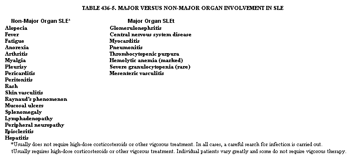
The variable severity and extent of involvement in SLE dictate individualized treatment. It is helpful to divide problems into those of major organs, which therefore are life threatening, and those that are unpleasant but not life threatening (Table 436-5). The major exception to this division is a syndrome of acute toxic lupus observed primarily in precorticosteroid times: a young woman with high fever, serositis, rash, and arthritis might succumb to SLE in the absence of major organ involvement. This syndrome is usually responsive to therapy with corticosteroids in modest doses. Non-major-organ involvements are best handled with symptomatic therapy: the less medicine the better. Hydroxychloroquine (200 to 600 mg per day) is effective for skin involvement; it also may help treat arthritis and other manifestations. Nonsteroidal anti-inflammatory drugs (NSAIDs) such as aspirin and ibuprofen are useful for arthritis, serositis, and fever. Some patients tolerate one NSAID better than another-bizarre neurologic reactions may occur in SLE patients receiving ibuprofen; liver enzyme abnormalities may follow aspirin treatment; gastrointestinal tolerance varies. The combination of hydroxychloroquine and NSAID may be sufficient. The addition of low doses of corticosteroids may be necessary. Initial every-other-day therapy may not be possible; however, a subsequent switch to alternate-day treatment reduces steroid-induced side effects. In patients with continued disease activity, symptoms may be prominent every other day, necessitating return to daily steroids. Even in the face of corticosteroid therapy, NSAID and hydroxychloroquine may add substantial benefit and allow a lower steroid dosage. Fevers occurring in spite of daily corticosteroids may respond to NSAIDs. Indomethacin may be especially effective in pericarditis. The management of major organ involvement is usually directed at preservation of function and prevention of organ
