|
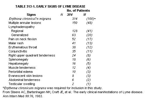
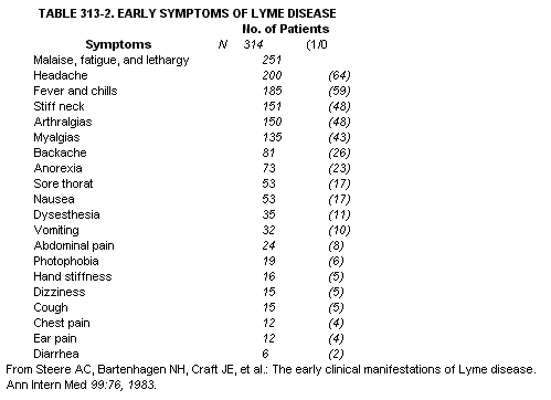
Lyme disease is a tick-borne inflammatory disorder caused by a newly recognized spirochete, Borrelia burgdorferi. Its clinical hallmark is an early expanding skin lesion, erythema chronicum migrans (ECM), which may be followed weeks to months later by neurologic, cardiac, or joint abnormalities. Symptoms may refer to any one of these four systems alone or in combination. Foci of Lyme disease are widely distributed within the United States and Europe "Lyme arthritis" was recognized in November 1975 because of unusual geographic clustering of children with inflammatory arthropathy in the region of Lyme, Connecticut. It soon became clear that this was a multisystem disorder (Lyme disease) occurring at any age, in both sexes, and often preceded by a characteristic expanding skin lesion, erythema chronicum migrans (ECM). In Europe ECM had been associated with the bite of the sheep tick, Ixodes ricinus, and with tick-borne meningopolyneuritis. In the Lyme region, a closely related deer tick, Ixodes dammini, was implicated as the principal disease vector on epiderniologic grounds. In 1982, Burgdorfer and associates isolated a spirochete from Ixodes dammini and linked it serologically to patients with Lyme disease. It was soon recovered from patient specimens DISTRIBUTION AND EPIDEMIOLOGY. Lyme disease is widespread. In the United States there are three distinct foci: the Northeast from Massachusetts to Maryland, the Midwest in Wisconsin and Minnesota, and the West in California, southern Oregon, and western Nevada. However, the illness has been reported in over one half of the states, as well as throughout Europe and in Australia. The earliest known cases in the United States occurred on Cape Cod in 1962 and in Lyme, Connecticut, in 1965; cases now number in the thousands. Disease can occur at any age and in either sex. Onset of illness is generally between May 1 and November 30, with the peak in June and July. The primary vectors of Lyme disease are tiny ixodid ticks. Major foci of disease correspond to the distribution of 1. dammini (Northeast and Midwest), I. pacificus (West), and I. ricinus (Europe), but other vectors, including the Lone Star tick, Amblyomma americanum, are likely in some areas. In one United States study, 31 per cent of 314 patients recalled a tick bite at the skin site where ECM developed days to weeks later. The six ticks that were saved were invariably nymphal I. dammini, whose peak questing period is May through July and which seems to be the stage primarily responsible for transmission of disease. Preferred hosts for I. dammini nymphs are white-footed mice and, for adults, white-tailed deer, in whose fur they may survive the winter. B. burgdorferi has been recovered from all three major ixodid tick vectors; in some areas, its prevalence in 1. dammini is as high as 60 per cent (compared with I. pacificus, ~ 1 per cent). The organism has been isolated, or specific antibody found, in blood and tissues of a wide variety of large and small animals, including domestic dogs and birds. Indiscriminate feeding on a variety of animals by immature L dammint may favor the spread of infection. PATHOGENESIS. Recovery of B. burgdorferi is straightforward from the tick (see above) but difficult from patients, perhaps because of a relative paucity of organisms in specimens of tissue and fluids from the latter. Nevertheless, rare positive cultures are reported at all stages of the illness-from blood (early), erythema chronicum migrans, meningitic cerebrospinal fluid, joint fluid, and even a late skin lesion, acrodermatitis chronica atrophicans, that had been present for 10 years. Spirochetes have been identified by silver stain or by immunofluorescence in some histologic sections of ECM and rarely of secondary annular lesions, synovium, brain, eye, and heart. From these data, combined with clinical (see below) and epiderniologic features of Lyme disease, the following pathogenetic sequence is likely. B. burgdorferi is transmitted to the skin of the host via the tick vector. After an incubation period of 3 to 32 days, the organism migrates outward in the skin (ECM), spreads in lymph (regional adenopathy), or disseminates in blood to organs (e.g., central nervous system and presumably liver and spleen) or other skin sites (secondary annular lesions; see below). Although organisms are hard to find in later stages of Lyme disease, it is likely that persistent live spirochetes are driving the illness throughout its course. Evidence for this interpretation includes the responsiveness of many patients to antibiotics, the rare sightings of spirochetes in affected tissues, and an expansion of the antibody response to additional spirochetal antigens over time. Lyme disease is associated with characteristic immune abnormalities. At disease onset (ECM), almost all patients have evidence of circulating immune complexes. At that time, the findings of elevated serum immunoglobulin M (IgM) levels and cryoglobulins containing IgM predict subsequent nervous system, heart, or joint involvement-i.e., early humoral findings have prognostic significance. Serial determinations of serum IgM are often the single most helpful laboratory indicator of disease activity. These abnormalities tend to persist during neurologic or cardiac involvement. Later in the illness, when arthritis is present, serum IgM levels are more often normal. By then, immune complexes are usually lacking in serum but are present uniformly in joint fluid. Mononuclear cells from peripheral blood increase their antigen-specific proliferative response as the disease progresses, but the greatest reactivity to antigen is seen in cells from inflamed joints. Adjacent to that joint fluid, one sees on biopsy a proliferative synovium often replete with lymphocytes and plasma cells that are presumably capable of producing immunoglobulin locally. Thus, an initially disseminated, immune-mediated inflammatory disorder becomes localized in some patients and propagated in joints. CLINICAL CHARACTERISTICS. Lyme disease is conveniently divided into three clinical stages, but the stages may overlap and most patients do not exhibit all of them. The illness usually begins with ECM and associated symptoms (stage 1), sometimes followed weeks to months later by neurologic or cardiac abnormalities (stage 2) and weeks to years later by arthritis (stage 3). Chronic neurologic and skin involvement may also occur years after onset. Early Manifestations Erythema chronicum migrans, the unique clinical marker for Lyme disease, begins as a red macule or papule at the site where the tick vector, usually long gone, had engorged. As the area of redness expands to 15 cm or so (range, 3 to 68 cm), there is usually partial central clearing. The outer borders are red, generally flat, and without scaling. The centers are occasionally red and indurated, even vesicular or necrotic. Variations may occurmultiple rings, for example. The thigh, groin, and axilla are particularly common sites. The lesion is warm to touch, but not often sore, and is easily missed if out of sight. Routine histologic findings are nonspecific: a heavy dermal infiltrate of monortuclear cells, without epidermal change except at the site of the tick bite. Within days of onset of ECM, one half of United States patients develop multiple annular secondary lesions. They resemble ECM itself but are generally smaller, migrate less, and lack indurated centers; they are not associated with the sites of previous tick bites. Individual lesions may come and go, and their borders sometimes merge. Other occasional skin lesions are noted in Table 313-1. In addition, benign lymphocytorna cutis has been reported in Europe. Erythema chronicum migrans and secondary lesions fade in three to four weeks (range, I day to 14 months). They may recur. malaise and fatigue, headache, fever and chills, myalgia, and arthralgia (Table 313-2). Some patients have evidence of meningeal irritation or mild encephalopathy-for example, episodic attacks of excruciating headache and neck pain, stiffness, or pressure-but typically lasting only for hours at this stage of the illness, and without spinal fluid pleocytosis or objective neurologic deficit. Except for fatigue and lethargy, which are often constant, the early signs and symptoms are typically intermittent and changing. For example, a patient may have meningitic attacks for several days, a few days of improvement, and then the onset of migratory musculoskeletal pain. This last may involve joints (generally without swelling), tendons, bursa, muscle, and bone. The pain tends to affect only one or two sites at a time and to last a few hours to several days in a given location. The various associated symptoms may occur several days before ECM (or without it) and last for months (especially fatigue and lethargy) after the skin lesions have disappeared. Skin involvement is often accompanied by flulike symptoms. Later Manifestations NEUROLOGIC INVOLVEMENT. Within several weeks to months of the onset of illness, about 15 per cent of patients develop frank neurologic abnormalities, including meningitis, encephalitis, chorea, cranial neuritis (including bilateral facial palsy), motor and sensory radiculoneuritis, or mononeuritis multiplex, in various combinations. The usual pattern is fluctuating meningoencephalitis with super_ imposed cranial nerve (particularly facial) palsy and peripheral radiculoneuropathy, but Bell's palsy may occur alone. By now, patients with meningitic symptoms have a lymphocytic pleocytosis (about 100 cells per cubic millimeter) in cerebrospinal fluid and sometimes diffuse slowing on electroencephalogram. However, the neck is rarely stiff except on extreme flexion; Kernig's and Brudzinski's signs are absent. Neurologic abnormalities typically last for months but usually resolve completely (late neurologic complications are noted below). CARDIAC INVOLVEMENT. Also within weeks to months (If onset, about 8 per cent of patients develop cardiac involvement. The commonest abnormality is fluctuating degrees of atrioventricular block (first-degree, Wenckebach, or complete heart block). Some patients have evidence of more diffuse cardiac involvement, including electrocardiographic changes compatible with acute myopericarditis, radionuclide evidence of mild left ventricular dysfunction, or, rarely, cardiornegaly, None has had heart murmurs. Cardiac involvement is usually brief (three days to six weeks), but it may recur. ARTHRITIS. From weeks to as long as two years after the onset of illness, about 60 per cent of patients develop frank arthritis, usually characterized by intermittent attacks of asymmetric joint swelling and pain primarily in large joints, especially the knee, one or two joints at a time. Affected knees are commonly more swollen than painful, often hot, and rarely red; Baker's cysts may form and rupture early. However, both large and small joints may be affected, and a few patients have had symmetric polyarthritis. Attacks of arthritis, which generally last from weeks to months, typically recur for several years. Fatigue is common with active joint involvement, but fever or other systemic symptoms at this stage are unusual. Joint fluid white cell counts vary from 500 to 110,000 cells per cubic millimeter, with an average of about 25,000 cells per cubic millimeter, mostly polymorphonuclear leukocytes. Total protein ranges from 3 to 8 grams per deciliter. The C3 and C4 levels are generally greater than one-third, and glucose levels usually greater than two-thirds, that of serum. Rheumatoid factor and antinuclear antibody are absent. In about 10 per cent of patients with arthritis, involvement in large joints may become chronic, with parmus formation and erosion of cartilage and bone. Synovial biopsy findings may mimic those of rheumatoid arthritis: surface deposits of fibrin, villous hypertrophy, vascular proliferation, and a heavy infiltration of mononuclear cells. In addition, there may be an obliterative endarteritis and (rarely) demonstrable spirochetes. In one patient with chronic Lyme arthritis, synovium grown in tissue culture produced large amounts of collagenase and prostaglandin E, Thus, in Lyme disease the joint fluid cell counts, the immune reactants (except for rheumatoid factor), the synovial histology, the amounts of synovial enzymes released, and the resulting destruction of cartilage and bone may be similar to those in rheumatoid arthritis. Other late findings (years) associated with this infection include a chronic skin lesion--acrodermatitis chronica atrophicans-well known in Europe but still rare in the United States. One sees violaceous infiltrated plaques or nodules, especially on extensor surfaces, that eventually become atrophic. Late chronic neurologic disease includes transverse myelitis and demyelinating lesions of the central nervous system
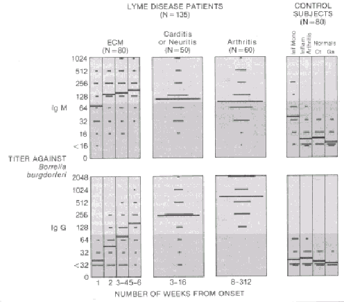
LABORATORY TEST RESULTS. Determination of specific antibody titers is currently the most helpful diagnostic test for Lyme disease. Culture of B. burgdorferi from patients permits definitive diagnosis but is rarely successful. Similarly, spirochetes are not often seen by direct examination of blood, plasma, plasma pellets, or skin transudate specimens of ECK and special tissue-staining techniques are low in yield and not readily available. In serum, specific IgM antibody titers against B. burgdorferi usually reach a peak between the third and sixth weeks after the onset of disease; specific immunoglobulin G (IgG) antibody titers rise more slowly and are generally highest months later when arthritis is present (Fig. 313-1). To date, no one with established Lyme arthritis has failed to have an elevated titer of specific IgG antibody. This finding makes antibody titers against B. burgdorferi particularly useful in differentiating Lyme disease from other rheumatic syndromes, especially when ECM is missed, forgotten, or absent. This antibody cross-reacts with other spirochetes, including Treponema pallidum, but patients with Lyme disease do not have positive VDRL test results. The most common nonspecific laboratory abnormalities, particularly early in the illness, are a high erythrocyte sedimentation rate, an elevated serum IgM level, or an increased serum glutarnic-oxaloacetic transaminase (SGOT) level. The enzyme levels generally return to normal within several weeks. Patients may be mildly anemic early in the illness and occasionally have elevated white cell counts with shifts to the left in the differential count. A few patients have had microscopic hernaturia, sometimes with mild proteinuria (dipstick); values for creatinine and blood urea nitrogen have been normal. Throughout the illness, serum C3 and C4 levels are generally normal or elevated. Rheumatoid factor and antinuclear antibodies are usually absent. DIFFERENTIAL DIAGNOSIS. Erythema chronicum migrans is the unique herald lesion of Lyme disease . When present in its classic form, there is little else that might be confused with it. However, some patients are not aware of having had ECM, and in others, its appearance is not always characteristic. Secondary lesions might suggest erythema multiforme, but blistering, mucosal lesions, and involvement of the palms and soles are not features of Lyme disease. Malar rash may suggest systemic lupus erythematosus; an urticarial rash, hepatitis B infection or serum sickness. Evanescent blotches and circles may resemble erythema marginatum, but those of Lyme disease do not expand. Early flulike symptoms may be misleading, especially when erythema chronicum migrans is absent or missed or is not the first manifestation. Severe headache and stiff neck may resemble aseptic meningitis; abdominal symptoms, hepatitis; and generalized tender lymphadenopathy and splenomegaly, infectious mononucleosis. As in the last infection, profound fatigue in Lyme disease may be a major and persistent complaint. In later stages, Lyme disease may mimic other immunemediated disorders. Like rheumatic fever, Lyme disease may be associated with sore throat followed by migratory polyarthritis and carditis, but without evidence of valvular involvement or of a preceding streptococcal infection. Migratory pain in tendons and joints may also suggest disseminated gonococcal disease. An isolated facial weakness may mimic Bell's palsy of other causes. Late neurologic involvement may suggest multiple sclerosis (transverse myelitis), Guillain-Barre' syndrome (symmetric peripheral neuropathy), primary psychosis, or brain tumor. In adults with Lyme arthritis, the large knee effusions can resemble those in Reiter's syndrome, and the occasional symmetric polyarthritis, that of rheumatoid arthritis. In children, the attacks of arthritis, although generally shorter, may be identical to those seen in the oligoarticular form of juvenile rheumatoid arthritis, but without iridocyclitis. FIGURE 313-1. Antibody titers against Borrelia burgdorferi
are shown in serum samples from 135 patients with different clinical
manifestations of Lyme disease, and from 80 control subjects with infectious
mononucleosis, inflammatory arthritis, or no disease (titers determined
by indirect immunofluorescence). The black bar shows the geometric mean
titer for each group; the pink shaded areas indicate the range of values
generally observed in control subjects. Note that all patients with
Lyme arthritis have elevated IgG antibody titers. (Adapted from Steere
AC, Grodzicki RL, Komblatt AN, et al: The spirochetal etiology of Lyme
disease. N Engl J Med 308:733-740, 1983.) TREATMENT Stage 1. In patients treated early with antibiotics, erythema chronicum migrans and its associated symptoms resolve in days, and subsequent major sequelae (myocarditis, meningoencephalitis, or recurrent arthritis) usually do not occur. For adults, therapy, in order of preference, includes oral tetracycline, 250 mg four times a day, phenoxymethyl penicillin, 500 mg four times a day, or erythromycin, 250 mg four times a day, each for 10 to'20 days, depending on the response. In children, phenoxymethyl penicillin, 50 mg per kilogram per day (not less than 1 gram or more than 2 grams per day), is given in divided doses for the same period, or, in cases of penicillin allergy, erythromycin, 30 mg per kilogram per day, in divided doses for 15 or 20 days. About 10 per cent of patients experience a Jarisch-Herxheimer-like reaction (higher fever, redder rash, or greater pain) during the first 24 hours of therapy. Whichever drug is given, nearly half of patients have brief (hours to days) recurrent episodes of headache or pain in joints, tendons, bursae, or muscle, often with lethargy, which may continue for extended periods. Such symptoms may represent undegraded antigen rather than persistent live spirochetes. Stage 2. For meningitis and cranial or peripheral neuropathies, intravenous penicillin G, 20 million units a day in six divided doses for ten days, is effective therapy. Headache, stiff neck, and radicular pain usually begin to subside by the second day of therapy and disappear by seven to ten days; motor deficits frequently require seven to eight weeks for complete recovery. Carditis also responds rapidly (in days) to this regimen. Prednisone, 40 to 60 mg a day in divided doses, may be added when a strong anti-inflammatory effect is important (e.g., persistent complete heart block). Stage 3. For established Lyme arthritis, even with unremitting involvement, parenteral penicillin is curative in some patients. During treatment, the affected joint should be at rest, and accumulated fluid removed by needle aspirations. In a double-blind, placebo-controlled trial, 7 of 20 patients given intramuscular benzathine penicillin, 2.4 million units weekly for three weeks, were cured (mean follow-up of 33 months), versus none of 20 control patients. This regimen provides low serum levels of penicillin for about six weeks. The high-dose intravenous regimen of penicillin G (above), which yields much higher serum levels over its ten-day course, cured 11 of 20 patients, including 2 in whom benzathine penicillin had failed. Optimal therapy for this problem and for the later neurologic complications of Lyme disease is not yet clear. Synovectomy may be useful for treatment failures. The infiltrative lesions of acrodermatitis chronica atrophicans are usually cured by three weeks of oral phenoxymethyl penicillin, 2 to 3 grams daily in divided doses. |


