|
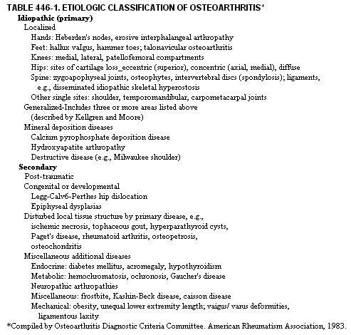
Osteoarthritis (OA) is a complex response of joint tissues to aging and to genetic and environmental factors, characterized by degeneration of cartilage, bony remodelling, and overgrowth of bone. Idiopathic osteoarthritis refers to the common variety encountered during aging that is unrelated to known systemic or local diseases and includes certain hereditary and erosive subsets. Secondary osteoarthritis refers to the form indistinguishable from the idiopathic (primary) type on a pathologic basis but clearly provoked by antecedent events, such as an inflammatory, metabolic, endocrine, developmental, or heritable connective tissue disorder (Table 446-1). Effects of a macrotrauma, repeated microtrauma, or prolonged immobilization on normal joints may dispose the joints to secondary osteoarthritis. PATHOLOGY. Minor cartilage softening in non-weightbearing sites and hypertrophic bony changes may persist a lifetime without producing symptoms. Osteoarthritis depends on development of progressively deepening clefts and erosions typically in weight-bearing sites. The disease advances over a period of years but rarely reaches the level of severity seen in rheumatoid arthritis; i.e., there is rarely joint fusion or parmus formation, and major subluxations are uncommon. When bony hypertrophy estimated by roentgenographic changes is used as a criterion, the majority of the population over 50 years of age is afflicted with osteoarthritis. By the eighth decade there is evidence of disease in 90 per cent of persons. It is the leading cause of joint pain and related disablements in middle-aged and elderly patients. The earliest histologic changes may be documented in the surface, subsurface, and deep zones of articular cartilage. These changes include loss of staining reactions for proteoglycans, with areas of cell injury, or loss followed by proliferation. On electron microscopic views, lipid accumulations, reduced cartilage collagen fibril size, edema, and surface irregularities are found. Clefts, microcysts, and erosions arise at the site of these changes. Aggressive lesions consist usually of vertical clefts in cartilage, which progress to deep erosions and exposure of subchondral bone. Bony thickening, eburnation, cysts, and bone-on-bone contact across the joint surface typify end-stage disease. ETIOLOGY. The most accepted premise is that primary changes in articular cartilage underlie development of osteoarthritis. Nevertheless, in a substantial subset of patients, biomechanical deficiencies arising from dysplasias of major or minor nature are causative. Similar biornechanical deficiencies may arise related to adolescent and adult remodelling of bones and abnormal distribution of weight-bearing forces (Table 446-1). Repeated industrial or sports-invoked macro- and microtraumatic events may produce excessive wear and hypertrophic remodelling responses. Evidence has been obtained for reduced biornaterial properties of cartilage as a function of aging and for possible metabolic disturbances in cartilage metabolism, as in diabetes mellitus, acromegaly, and ochronosis. The subchondral bone table is disturbed by tissue remodelling early in the disease or by such afflictions as Paget's disease or hyperparathyroidism with subchondral cysts. Hyperlaxity of ligaments per se or as part of certain overt (heritable) disorders of connective tissue can lead to osteoarthritis. PATHOGENESIS. As a result of a multiplicity of etiol , ogic factors, an apparent final common pathway of disease expression involves breakdown of cartilage both by direct physical injury and by enzymatic degradation resulting from injury to chondrocytes and indirectly by subchondral bone stiffening from remodelling. Most important in this context is injury of the collagen network or framework that holds together articular cartilage. This network retains in a semidehydrated conformed state the abundant, intensely hydrophilic, charged, proteoglycan macromolecules. The latter exert an osmotic pressure of several atmospheres against the network. Ungluing or cleavage of the collagen network by maldistributed or excessive weight-bearing forces, or degradation of the network by enzymes elaborated by injured cartilage cells may occur. This response to various precipitating events leads to loss of essential elastic properties In early osteoarthritis, repair responses by local chondrocytes are usually of a poor quality, leading to almost no replacement of lost tissue. From advanced erosions penetrating the marrow, tissue repair is more effective and consists of mixtures of fibro- and hyaline cartilage. Normal rugged biornaterial properties are never recovered by the repair cartilage. As degeneration proceeds, wear particles and matrix degradation products are released from both original and repair cartilages. These fragments are carried to the synovial lining membrane where a phagocytic response engenders low-grade inflammation and synovial effusion, proliferation of synovial cells, and thickening of the synovial membranes. Much evidence has accumulated that membrane-engendered inflammatory factors amplify cartilage breakdown. Several biochemical abnormalities have been noted in osteoarthritic cartilage: increased water content, decreased aggregation and content of proteoglycans, decreased chain length and altered profiles of glycosaminoglycans, and increased proteolytic enzyme levels. CLINICAL MANIFESTATIONS. The clinical presentation may be divided into early and late stages. Throughout these stages, there is deep, aching pain in the afflicted joints, morning stiffness of short duration, and variable joint thickening and effusion. Early stages are dominated by pain on motion with stiffness, night pain, and responsiveness to antiinflammatory medication. The late stages are dominated by joint instability, predominance of pain at rest accentuated on weight-bearing, and failure of responsiveness to anti-inflammatory agents. The present description of clinical features is developed largely on an anatomic basis inasmuch as signs and symptoms reflect regional patterns of involvement. Roentgenographic and laboratory work-up and treatment are discussed later.
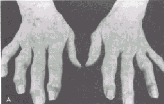
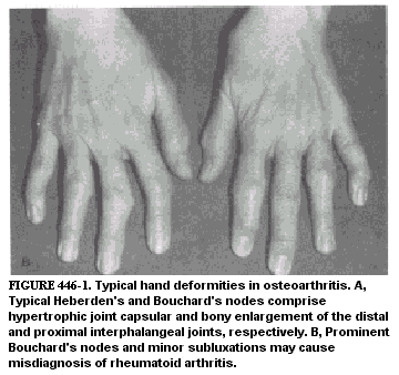
Hands. Heberden's nodes refer to the osteoarthritic disfigurements of distal interphalangeal joints, and Bouchard's nodes signify equivalent lesions of the proximal interphalangeal joints of the hands (Fig. 446-1). Early Heberden's nodes have a soft consistency and may be associated with prominent inflammatory signs. In the chronic stage, they are characterized by bony enlargement and angular deformities with variable symptoms. Heredity and sex, in addition to microtrauma, are prominently involved in the development of Heberden's nodes, which are more common in women at menopause or late middle age. The only other common hand lesion involves the first carpometacarpal joint. Such lesions are associated with pain in the radial side of the wrist, intensified by physical activities such as golf, tennis, and knitting
Knees. The commonest source of major disability in osteoarthritis is from knee involvement. At any one time, heat, synovial thickening, or effusions have been documented in at least 50 per cent of cases. Elicitation of crepitus, which persists on repeated flexion and extension of the knee, bony marginal overgrowth, mediolateral instability (in the late stages), and synovial effusion are important diagnostic aids. Degenerative changes are usually more prominent in the medial compartment of the knee, leading to varus (bowleg) deformities. Developmental defects, i.e., knock-knee or bowleg deformity, predispose to CA. Degenerative alteration of the patellofemoral joint is termed chondromalacia patellae. This syndrome of mild effusion and knee pain is usually associated with trauma and occurs predominantly in young adults. It is usually preceded by developmental biornechanical disturbances influencing knee flexion. There is often spontaneous remission of symptoms, but some cases progress to irreversible patellofemoral osteoarthritis. Malum Coxae Senilis (Coxarthrosis). Clinical manifestations of primary hip joint disease appear usually in late middle or old age. Perhaps one third or more of cases arise from acetabular dysplasia, as well as growth or maturational disturbances in the femoral neck and head. Altered bone growth as well as developmental thickening at the zenith of the acetabulum may be causative in less than 5 per cent of cases. Beyond these factors, there is a background of adult bone remodelling and altered joint incongruity, which may further compromise normal weight-bearing patterns and chondrocyte nutrition. In addition, a variety of acquired disorders such as rheumatoid arthritis and ischernic necrosis of the femoral head are important etiologically. Groin pain on weight-bearing or motion is a dominant symptom and is usually referred to the anterior aspect of the thigh above the knee. Over a period of months or a few years, invalidism from severely restricted mobility and pain is a common outcome of untreated disease. Spinal Osteoarthritis (Including Herniated Disc Syndrome). Throughout the spine, weight-bearing compressive forces are largely supported by one set of articulations-the intervertebral discs. These are elastic organs similar to articular cartilage in respect to the fact that they depend on properties of the semi-dehydrated proteoglycan molecules. A high osmotic pressure at rest results from proteoglycan confinement by cartilage end-plates in two dimensions and the annulus fibro sus in the third. An additional important elastic component is provided by the annulus fibrosus. Rotary motions in the back and neck depend upon the zygoapophyseal joints. All of these joints undergo osteoarthritic changes almost identical in nature to those of the peripheral joints (see Ch. 448 for specific clinical features). Ordinarily, the former joints protect the latter against severe torsional trauma; under certain conditions, especially flexion of the lumbosacral spine, the rotary joints are of much less protectional value, and annular tears may occur under these (and several other) conditions. Resultant displacement of discal products into the spinal foramen adjacent to nerve rootlets and/or spinal canal occurs, depending on the conditions of damage. Injury of these structures both by mechanical trauma and by activated inflammatory pathways constitutes one of several causes of neural dysfunction leading to symptoms. The relationship of the posterior zygoapophyseal joints in the cervical, thoracic, and lumbar spines to their respective nerve roots as they traverse the intervertebral foramina, and the proximity of an additional set of joints of Luschka in the cervical spine (segments C2 to C7) have similar importance because of potential damage to nerves by inflammation secondary to mechanical irritation. Notably, symptoms of osteoarthritis in the cervical spine depend upon the neural segment involved. Pain, aggravated by motion, often radiates into the supraclavicular and upper trapezius regions, as well as the occiput and distal upper extremities. Overgrowth of bone in either the cervical or lumbar spine can cause narrowing of the spinal canal and encroachment on the spinal cord rather than the nerve roots. In the neck, a myelopathy of a painless nature may result. Constriction of the spinal cord by surrounding bone, disc, or ligamentous thickening leads to the syndrome of spinal stenosis most common in the lumbosacral spine. Neurogenic claudication pain (resembling vascular claudication) is an important symptom in this condition and must be differentiated from vascular insufficiency (see Ch. 448 for management of discogenic claudication). Diffuse Idiopathic Skeletal Hyperostosis. This is characterized by a flowing ligamentous calcification along the anterolateral aspects of vertebral bodies. Symptornatology is variable and focused in the spine or multiple tendon osseous junctions (e.g., at the heel); ankylosis of apophyseal and sacroiliac joints is absent. The thoracic spine is most often affected without intervertebral disc narrowing. LABORATORY FINDINGS. There are no specific abnormalities in osteoarthritis. The sedimentation rate is usually within normal limits, synovial fluid is clear and exhibits a normal range of viscosity, and there is a negative mucin clot test result. Leukocyte counts in synovial fluid generally vary from 150 to 1500 per cu mm; wear particles including whole fragments containing proteoglycans and collagen fibers as well as mineral particles are often identified in the fluid. ROENTGENOGRAPHIC FEATURES. There is usually narrowing of the radiolucent interosseous joint space resulting from destruction of articular cartilage. Bony cysts varying in size may be seen in subchondral or denuded bone, which may be densely sclerotic. Osteophyte formation at the margins of affected joints is the basis for the most striking roentgenographic findings. Degeneration of lumbar and cervical intervertebral discs results in narrowing of the interspaces. A vacuum sign or marked translucency in the disc may be seen. Evidence for disc degeneration is usually documented in anteroposterior and lateral roentgenograms. For visualization of osteophytes blocking foramina, particularly in the cervical spine, oblique views are mandatory. Oblique views are also valuable for defining bony sclerosis and joint-space narrowing of the zygoapophyseal joints of the lumbar spine. DIFFERENTIAL DIAGNOSIS. Osteoarthritis and rheumatoid arthritis are readily distinguished in terms of their usual clinical presentation. The latter is generally associated with prominent signs of joint inflammation, characteristically afflicting the hands and wrists symmetrically, especially the metacarpophalangeal joints. These joints almost never are affected in osteoarthritis. Differentiation of these disorders is more complicated when seronegative rheumatoid arthritis involves only (or predominantly) the lower extremities. The presence of a normal erythrocyte sedimentation rate, negative serum rheumatoid factor test result, and minimal synovial fluid change supports the diagnosis of osteoarthritis. Despite severe deformities, occasionally seen with Heberden's and Bouchard's nodes, the lack of uInar drift and metacarpophalangeal and diffuse wrist involvement help to rule out rheumatoid arthritis. Erosive osteoarthritis characteristically shows bony destruction and inflammatory changes in the proximal and distal interphalangeal joints but not in the metacarpophalangeal joints. Secondary osteoarthritis must be considered in the presence of joint hypermobility, chondrocalcinosis, heritable disorders such as the Ehlers-Danlos syndrome, mechanical derangements of the joints, metabolic bone disorders, ochronosis, neuropathies, and hemochromatosis. Spinal involvement in osteoarthritis is distinctly different from that in ankylosing spondylitis; the latter predominantly afflicts young men and has characteristic and distinctive roentgenographic features involving sacroiliac sclerosis and fusion, calcification and ossification of the annulus fibrosus and adjacent paravertebral ligaments, and formation of bridging syndesmophytes (bamboo spine). TREATMENT. Although treatment depends in large measure on the site and severity of joint involvement, the outlook with a multidisciplinary long-term management program is relatively optimistic for functional restoration and symptomatic improvement. Early disease with signs of mild to moderate inflammation but without joint instability can usually be managed successfully with a combination of measures: (1) Relief of pain with mild analgesics (e.g., acetaminophen 500 mg three or four times a day) or nonsteroidal anti-inflammatory agents (e.g., aspirin 2400 to 3600 mg daily) or both. Indomethacin, ibuprofen, naproxen, fenoprofen, piroxicam, sulindac, and tolmetin are alternative agents, advantageous as substitutes for aspirin. Where possible, intermittent rather than continuous usage is encouraged to avoid gastrointestinal side effects, especially acid peptic disease. (2) Revision of daily schedule of activities, increased joint rest, and selected avoidance of activities unfavorable to the symptomatic joints. (3) Protection of joints with relevant devices, i.e., splints, crutches, walkers, canes, etc. (4) Use of weight-reducing diets. (5) Diazepam, 5 mg three to four times daily or another suitable agent may be used sparingly for acute episodes of muscle spasm. (6) Application of moist heat or cold packs may help. (7) Symptomatic response, in refractory cases, to intra-articular or paraarticular injections of small amounts of corticosteroid at infrequent intervals is useful. (8) Once pain and muscle spasm have been relieved, a formalized program of physical therapy followed by a prolonged home exercise program is often recommended in the hope of retarding further joint deterioration. In the case of cervical osteoarthritis, hyperextension and hyperflexion should be avoided. The patient should sleep flat on one pillow and employ intermittent traction devices available for home use. A cervical collar restricts motion and minimizes pain. (See Ch. 448 for treatment of osteoarthritis in the lumbar spine.) The principal anatomic regions that are most benefited by orthopedic surgery are the knee, hip, and spine. Several surgical procedures are appropriate for patients with severe hip involvement, including osteotomy, mold arthroplasty, total joint replacement, and arthrodesis. For the knee, debridement, either through an arthroscope or via open surgery, osteotomy, and a variety of partial or complete arthroplasties are used for treatment. Tibial or femoral osteotomies may be of long-term benefit by realigning weight-bearing forces, but considerable follow-up rehabilitation is required. Otherwise, joint replacement is the treatment of choice for many cases of advanced osteoarthritis of the knees characterized by intractable pain, loss of function, instability, or all three. Some indications for spinal surgery are: |


