ENTRANCE | HOME | 1 | 2 | 3 | 4 | LINKS | FUN STUFF | BULLETIN BOARD | BOOK STORE | DISEASES | SEARCH
Ralph Snyderman
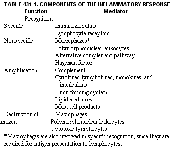
Immunologic processes mediate the localization and destruction of substances that, if disseminated, could disrupt the host's complex internal milieu. The immune system has several unique features that permit it to combat microbial invasion and provide resistance against the spread of cancer. Unlike *other tissues, the immune system consists not only of fixed structures (i.e., thymus, spleen, and lymph nodes) but also of motile cells that wander throughout the body, performing surveillance. It is also the only tissue able to destroy other components of the host. Both the protective and destructive abilities of immunologic processes relate largely to their inflammatory potential. Understanding how inflammation is initiated is thus essential for understanding the mechanisms of immunologically mediated resistance and for comprehending how tissue destruction occurs in the rheumatic disorders. To fulfill its function of host defense, the immune system must differentiate self from nonself and then rapidly destroy substances recognized as nonself. A progression of immunologic recognition, amplification of the immune reaction, accumulation of inflammatory cells, and finally destruction of the inciting agent is an ongoing subclinical process. Inflammatory reactions can be initiated by either specific or nonspecific medns (Table 431-1). Recognition of unique determinants (epitopes) on antigens by antibodies or by receptors on lymp~ocytes initiates immunologically mediated inflammation. Nonspecific recognition can be initiated by components of the immune system that bind to materials based on their charge, hydrophobicity, or lectin composition. Nonspecific recognition is mediated in part by phagocytic cells, such as polymorphonuclear leukocytes and macrophages, as well as by C3b, an initiator ofthe alternative pathway of the complement system (see Ch. 418), and by Hageman factor.
Following recognition of nonself, amplification systems are activated and lead to the production of mediators of inflammation. The type of amplifier involved depends upon the recognition component and the nature and location of the inciting material. Amplification components of the immune system such as complement cleavage products, cytokines, and other phlogistic factors magnify the initial response to nonself Infla atorv reactions can also be initiated bv non-immunologic means. For example, inflammation following tissue necrosis results from the direct cleavage of complement components by lysosomal proteases released by injured cells. This phenomenon may play a role in extending cardiac: tissue damage following myocardial infarction. In gout or pseudogout, inflammation follows the phagocytosis, by polymorphonuclear leukocytes, of monosodium urate or calcium pyrophosphate dihydrate crystals. Ingestion of these agents by leukocytes leads to the release of lysosomal hydrolases as well as the production of chemotactic factors, which attract other inflammatory cells into the joint.
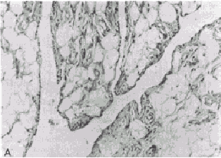
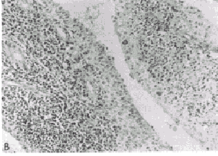
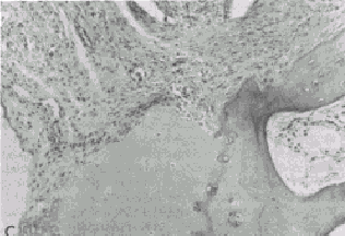
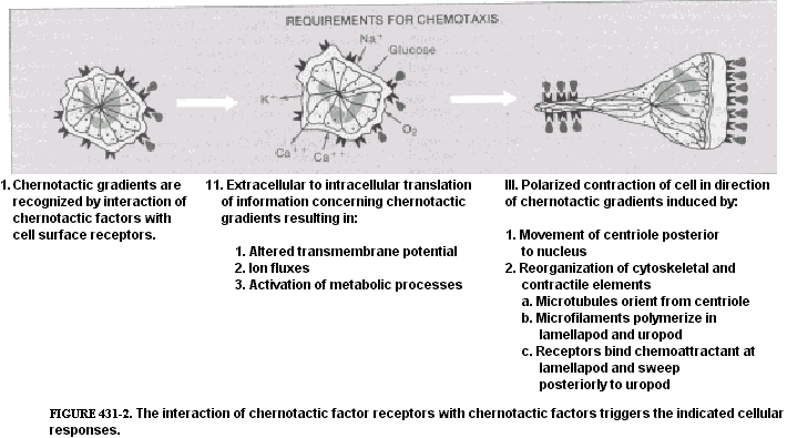
Regardless of the type of inflammatory response, the accumulation of granulocytes and macrophages can result in phagocytosis and degradation of the material that initiated the inflammatory event. The factors that determine whether an inflammatory response will be protective or destructive depend in part upon the nature and location of the inciting agent, its quantity, digestibility, the genetic makeup, and the immunoregulatory competency of the host. In general, when the antigen or other inciting agent is rapidly disposed of, the inflammatory process is self-limited. When antigen persists or is excessive in amount, the inflammatory response can be locally destructive and become clinically apparent. RHEUMATOID SYNOVITIS AS A MODEL OF INFLAMMATORY-MEDIATED TISSUE DESTRUCTION A chronic, locally destructive inflammatory reaction in humans is exemplified by the synovitis present in some connective tissue disorders. The prototype disease is rheumatoid arthritis. The diarthrodial joint has several features that influence inflammatory processes that occur there. The synovial membrane is highly vascular and lines all intra-articular structures, except for cartilage. The synovial lining is devoid of a basement membrane and thus permits relatively free diffusion of soluble substances. Moreover, the synovium lines a closed cavity, the joint space; therefore any reactive materials gaining entrance to the joint space are difficult to remove. Normal synovium is a thin layer of tissue whose lining is composed of two principal cell types supported by a loose connective tissue stroma (Fig. 431-1A). The Type A cell is rich in surface pseudopodia, and its cytoplasm has many lysosomes and prominent Golgi complexes but little rough endoplasmic reticulum. Type A synoviocytes have many characteristics of macrophages and are phagocytic, and ultrastructural studies have implied a secretory role for these cells as well. The Type B synoviocyte exhibits prominent rough endoplasmic reticulum but few vacuoles, lysosomes, or cell processes. This cell is primarily a secretory cell, hyaluronic acid being an important product. Synoviocytes may, however, be multipotential, with their morphology reflecting the net result of stimuli present in the local milieu. Beneath the synovial membrane there are collagen fibrils, fatty tissue, and an extensive capillary network. Fibroblasts in this area produce Types I and III collagen. Lastly, the dense fibrous joint capsule provides support for the synovial lining membrane and separates the articular space from surrounding structures. Rheumatoid synovitis exhibits three main components: inflammation, proliferation, and infiltration. In early stages, both Types A and B cells proliferate and increase in size. Fibrin deposits are frequently present on the inner synovial lining. Polymorphonuclear leukocytes predominate in the synovial fluid exudate, but cells are seen as infiltrates only in the superficial synovial layer. The supporting stroma beneath the lining cell layer becomes edematous and develops an increase in the number of small blood vessels. Concurrently, there are focal accumulations of inflammatory cells consisting of lymphocytes, plasma cells, macrophages, and occasionally mast cells. If the inflammatory synovitis persists, a proliferative lesion develops, characterized by synovial membrane thickening and projection of villous formations into the articular cavity (Fig. 431-1B). there is a concomitant increase in supporting connective tissue, small blood vessels, mononuclear cell infiltrates, and occasional multinucleated giant cells, as well as increased numbers of undifferentiated mesenchyme-like cells that have both phagocytic and synthetic potential. Clusters of poorly differentiated synovial cells may be found invading cartilage and subchondral bone. As the proliferative lesions progress, fibrous-mesenchymal tissue (pannus) begins to invade and replace cartilage and bone at the periphery of the synovial reflection (Fig. 431-1C). The invasion of cartilage, subchondral bone, and tendon by inflammatory synovial tissue results in collagen destruction, degradation of proteoglycans in the cartilage matrix, and bony resorption. MEDIATORS OF INFLAMMATION THAT PARTICIPATE IN THE RHEUMATIC DISEASES Inflammatory reactions result from the local production of a number of mediators derived from humoral or cellular sources. A complex interplay of activation and suppression mechanisms modulates the type and magnitude of the inflammatory response. LIPID MEDIATORS. Phospholipids are major constituents of cell membranes, including those of leukocytes and platelets, and are subject to degradation by cellular phospholipases under certain conditions such as exposure of cells to inflammatory or noxious stimuli. Cleavage of phospholipids results in the release of arachidonic acid, which can be further metabolized into a number of biologically potent mediators and modulators of inflammation. Prostaglandins (PG) are synthesized from arachidonic acid following the action of the enzyme cyclo-oxygenase, which forms PGG, which is then reduced to PGH,. Depending upon the isomerase enzymes present in the particular tissue, prostaglandins (PGE,, PGF,,), thromboxanes, or prostacyclins will be formed. Leukocytes and explants of rheumatoid synovia produce predominantly PGE,, platelets produce thromboxane A, and endothelial cells produce prostacyclin. Thromboxanes are vasoconstrictors, whereas prostacyclins are vasodilators. PGE, appears to modulate a number of inflammatory events. It enhances vascular permeability, is pyrogenic, and increases sensitivity to pain. PGE, also stimulates formation of cAMP in many types of inflammatory cells and thereby suppresses a number of immunologic responses, including release of mediators from mast cells, lymphocyte blastogenesis, and lymphocyte-mediated cytotoxic reactions. An important source of PGE, in immunologic reactions is the macrophage. Supernatant fluids from explants of rheumatoid synovium stimulate bone resorption by enhancing osteoclast activity and release of bone calcium. This phenomenon is probably mediated in large part by PG, since it is inhibitable by indomethacin, which blocks their formation. Arachidonic acid can also be metabolized into another class of biologically active derivatives by the enzyme lipoxygenase. The hydroxy-eicosatetraenoic acids (HETEs) and the derivatives of 5-hydroperoxy-eicosatetraenoic acid (termed leukotrienes) are examples of these arachicionic acid metabolites and are synthesized by granulocytes, macrophages, and basophils. 5,12-HETE, also termed leukotriene B, (LTB,), is a potent chernotactic factor, whereas leukotrienes C and D stimulate bronchoconstriction. LTB, has been identified in the synovial effusions of patients with rheumatoid arthritis and ankylosing spondylitis. Large amounts of LTB, are produced when granulocytes phagocytize monosodium urate crystals, and production of LTB, is inhibited by colchicine. This chemoattractant may, therefore, be important in the pathogenesis of gouty inflammation. Platelet-activating factors (PAF) are a group of acetyl-alkylglycerol ether analogues of phosphatidylcholine. PAF causes platelet aggregation and is a potent leukocyte activator and chemoattractant. Leukocytes produce PAF following their stimulation by inflammatory mediators. BIOLOGICALLY ACTIVE AMINES. Histamine and Serotonin. Histamine is derived from the decarboxylation of histidine by the enzyme L-histidine decarboxylase. The majority of histamine is stored in mast cells and basophils and is complexed with mucopolysaccharides such as heparin. Stimulation of mast cells and basophils by a number of mechanisms causes secretion of histamine. This agent has diverse biologic activities, including constricting smooth muscle, enhancing vascular permeability, depressing leukocyte chemotaxis, blocking T lymphocyte function, and depressing further histamine release from mast cells and basophils. Histamine thus may modulate both acute and chronic inflammatory responses. Serotonin (5-hydroxytryptamine) is produced by the decarboxylation of 5-hydroxytryptophan. More than 90 per cent of body stores of serotonin are found in the gastrointestinal tract and the central nervous system; the remainder is present in the dense granules of platelets, The biologic role of serotonin in inflammation is not well understood, but it enhances the chernotactic responses of leukocytes and increases fibroblast growth in vitro. It also stimulates collagen formation. BIOLOGICALLY ACTIVE PEPTIDES. Complement Cleavage Products. The complement (C) system functions as an important amplifier of inflammatory events initiated by immunoglobulins IgG and IgM as well as inflammatory reactions initiated by release of hydrolytic enzymes from traumatized cells or by leukocytes. The biology and biochemistry of this complex series of proteins are described in Ch. 418. Two complement cleavage products, C3a and C5a, derived from the third and fifth C components, respectively, are mediators of inflammation in rheumatic disorders such as rheumatoid arthritis and will thus be described in greater detail here. C3a: Enzymatic cleavage of the a. chain of C3 by the earlieracting C components or by other proteases releases C3a, a peptide consisting of 77 amino acids. C3a mediates a number of biologic responses, including smooth muscle contraction, vasodilatation, enhanced vascular permeability, the degranulation of mast cells and basophils, and the secretion of lysosomal enzymes by leukocytes. C3a also has immunoregulatory effects and suppresses humoral immune responses in vitro by affecting T lymphocytes. C3a is the most abundant of the C peptides released upon activation of C in serum. The COOH-terminal arginine of C3a is required for its biologic activity, and cleavage of this amino acid by a carboxypeptidase-B-like enzyme in serum renders the peptide inactive. C5a: C5a has a number of structural and biologic similarities to C3a. C5a consists of 74 amino acids, the COOH-terminal constituent also being arginine. C5a is derived from cleavage of the ot chain of C5. In addition to having all the biologic activities of C3a, C5a is also an extremely potent chemoattractant for polymorphonuclear leukocytes, monocytes, and macrophages. C5a is the major source of chernotactic activity generated in serum treated with immune complexes or endotoxin and is also an important source of chernotactic activity in rheumatoid synovial fluids. In contrast to C3a, C5a potentiates humoral immune responses in vitro. Cleavage of the terminal arginine of C5a by a serum carboxypeptidase-B markedly diminishes its biologic activity. Crystal-Induced Chemotactic Factors. Leukocytes accumulate in the-synovial fluid of individuals with gout or pseudogout following the ingestion by neutrophils of monosodium urate or calcium pyrophosphate dihydrate crystals, respectively. Incubation of neutrophils with these crystals in vitro results in the production by the cells of LTB, and a polypeptide chemoattractant (CCF) with a molecular weight of 8400. The production of these chemoattractants is blocked by colchicine. A mechanism by which colchicine abrogates acute gouty arthritis may be its ability to inhibit the synthesis of chemoattractants by neutrophils. Kinin-Forming System. An intimate association exists between the activation and regulation of the intrinsic clotting, fibrinolytic, and kinin-forming systems. Hageman factor (HF) (Factor XII of the clotting system) is central to the activation of all three systems. HF is activated nonspecifically by a number of agents, including exposure to crude preparations of collagen, vascular basement membranes, monosodium urate crystals, calcium pyrophosphate crystals, and endotoxin. Negatively charged surfaces also activate HF. Upon activation, HF (an 80,000-dalton P globulin) is cleaved, and its active form HF, initiates the conversion of Factor XI of the clotting pathway to XIa, and the conversion of prekallikrein to kallikrein. Kallikrein activates plasminogen, an enzyme important in fibrinolysis. Kallikrein also cleaves serum kininogen to form bradykinin, a nonapeptide with potent biologic activities. Cl esterase inhibitor (ClINH), a protein that inhibits activated Cl, is also an important inhibitor of HF, and kallikrein. Bradykinin and two other kinins produced by tissue kallikreins from kininogen induce smooth muscle contraction, increase vascular permeability, and induce pain. Cleavage of fibrinogen by plasmin results in production of a number of products, including fibrinopeptide B, which potentiates the action of bradykinin and has chemotactic activity. Interleukins and Other Cytokines. Interleukins are immunoregulatory molecules synthesized by mononuclear leukocytes. Stimulation of macrophages by antigens as well as by factors from lymphocytes initiates the secretion of interleukin 1 (IL-1), a 15,000-dalton peptide with diverse biologic activities. Macrophages are required for many of the activities of lymphocytes, and certain of the "helper" functions of macrophages are mediated by IL-1. IL-1 may be identical to a factor termed mononuclear cell factor (MCF), which stimulates synovial cells to produce collagenase. IL-1 may also be identical to leukocytic pyrogen. Tumor necrosis factor-a (TNF-(x) is a macrophage product with IL-l-like activities plus the ability to kill many types of tumor cells (see Ch. 257). Interleukin 2 (IL-2), previously termed T-cell growth factor, is a 15,000-dalton polypeptide produced by T lymphocytes, which stimulates the continuous proliferation of activated T lymphocytes in culture. Transforming-growth factor-P (TGFP) is a 25,000-dalton homodimeric protein found in platelets and produced by stimulated lymphocytes. TGF-P inhibits further lymphocyte division and regulates differentiation of many cell types including fibroblasts. lnterferon--~ (IFN--~) is a 50,000 dalton protein produced by stimulated lymphocytes. IFN-,y activates macrophages and synergizes with other immunomodulators. LYSOSOMAL ENZYMES. Lysosomal enzymes are contained in subcellular organelles termed lysosomal granules. These enzymes degrade complex macromolecules. Lysosomal granules also contain antimicrobial constituents such as myeloperoxidase and lactoferrin. Leukocytes contain several types of lysosomal granules, two of which (primary and secondary) are differentiated by their staining characteristics. Lysosomal enzymes digest antigens following phagocytosis. However, since these enzymes may also be released during phagocytosis or upon cell death, they can cause tissue destruction. Lysosomal proteases found in leukocytes include collagenase, elastase, cathepsin D, cathepsin G, and gelatinase. These enzymes are capable of destroying extracellular structures and may participate in mediating tissue injury in the rheumatic diseases. Cathepsin D cleaves cartilage proteoglycan, whereas granulocyte collagenase is active in cleaving Type I and, to a lesser degree, Type III collagen of bone, cartilage, and tendon. Substrates of granulocyte elastase include collagen crosslinkages and proteoglycans, as well as elastin components of blood vessels, ligaments, and cartilage. Lysosomal hydrolyases also produce mediators of inflammation through their direct action on C components such as C5. Leukocytic hydrolyases can liberate kinin from kininogen. Plasminogen activator, an enzyme that converts plasminogen to plasmin (which stimulates fibrinolysis) is found in both granulocyte and macrophage lysosomes. Rheumatoid synovial collagenase is usually present as an inactive lysosomal proenzyme that requires plasminogen activator for conversion to its active form. Regulation of tissue destructive potential of the lysosomal proteases is mediated by protease inhibitors such as a, macroglobulin and a, antiprotease. These antiproteases are present in serum and in synovial fluids and inhibit proteases by binding to them and covering their active sites. PHYSIOLOGIC MECHANISMS OF INFLAMMATORY CELL ACCUMULATION The accumulation of inflammatory cells at sites of antigen is central to the inflammatory process. Polymorphonuclear leukocytes and macrophages are motile cells that have many common physiologic characteristics. Both can perceive gradients of chemoattractant molecules and migrate directionally along such gradients. They also perform endocytosis (ingestion), secrete lysosomal enzymes, and generate superoxide anions (see Ch. 148). Polymorphonuclear leukocytes and macrophages have specific surface receptors that perceive C5a, CCF, and LTB, as well as synthetic polypeptide chemo tactic factors . Binding of chemoattractants to the surface of phagocytes results in orientation of the cells toward the source of the chemotactic gradient. The cells lose their round configuration and become triangular, with the base of the triangle facing toward the chemoattractant gradient (Fig. 431-2). This change in cell shape requires rearrangement of intracellular cytoskeletal elements. Microtubules provide a front-to-back polarization, whereas actin filaments accumulate at the front and back of the cells and provide the contractile forces required for movement. Attachment of leukocytes to the vascular endothelium and other surfaces is a requirement for cellular motility. Chemoattractants stimulate the appearance of several adhesive proteins on the surface of phagocytes. These proteins include the iC3b receptor (mac 1, mo 1), LFA 1, and glycoprotein 150/90. These proteins are heterodimers that serve a number of adhesive functions. They consist of individual ot-chains and a common P-subunit. Their appearance on the surface of phagocytes enhances the cells' binding to endothelial cells, tumor cells, and other surfaces. Deficiency of these proteins is associated with a severe defect of leukocyte function and a marked increase in susceptibility to infection. Chemotactic factors can also initiate other cellular responses by leukocytes, such as superoxide anion production and lysosomal enzyme secretion. The concentration of chernotactic factors required to initiate these latter processes is approximately 10-fold greater than that required for the induction of chemotaxis. Thus, release of potentially toxic products from the cells may not occur until they arrive at the inflammatory site where the concentration of chemoattractants is high. |
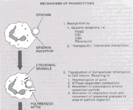
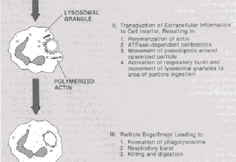
Chemoattractants activate leukocytes through their binding to cell surface receptors. Occupancy of chemoattractant receptors causes a guanine nucleotide regulatory (G) protein to become activated through the dissociation of GDP, followed by the binding of GTP. The activated G protein lowers the calcium concentration required to stimulate a membraneassociated phospholipase C, which in turn hydrolyzes a membrane phospholipid. termed phosphatidylinositol 4,5-bisphosphate. This hydrolysis leads to the production of two second messengers, the calcium mobilizer inositol 1,4,5-trisphosphate (IP,) and the protein kinase C activator, 1,2-diacylglycerol. When the dose of chemotactic factor is sufficient, protein kinase C becomes translocated from the cytosol to the plasma membrane of the phagocyte and activates the respiratory burst. Feedback inhibition of this activation pathway is mediated by transient cAMP elevation, which inhibits IP, production, as well as by protein kinase C, which uncouples the ability of the activated G protein to stimulate phospholipase C. Lymphocytes are also motile cells, but they do not respond to the same chernotactic factors as polymorphonuclear leukocytes and macrophages. The migration of lymphocytes is stimulated by specific antigens, mitogens, and other undefined factors produced by lymphocytes. Phagocytosis is initiated by binding of a particle to the surface of phagocytic cells. Particles carrying bound immunoglobulin or the complement fragments C3b or C3bi are more readily phagocytosed, since they bind to Fc and C3b receptors on both polymorphonuclear leukocytes and macro- phages. However, phagocytosis can occur in the absence of receptor involvement. Particle ingestion results from the envelopment and fusion of phagocytic cell membrane around the foreign material (Fig. 431-3). Intracellular lysosomes migrate to the phagocytic vesicle, fuse with it, and empty their contents, thus forming a phagolysosome. Within the phagolysosome, antigenic digestion and microbial killing generally occur. During phagocytosis, lysosomal hydrolases and toxic oxygen radicals may be released. IMMUNOLOGICALLY MEDIATED TISSUE INJURY Inflammatory responses can produce adverse reactions ranging from minor local tissue irritation to selective destruction of organs or even sudden death. The nature of immunologically mediated inflammatory responses depends upon the immunologic recognition component that identifies the antigen. Four general types of immunologically mediated inflammatory reactions have been defined (Table 431-2) * In clinical and experimental situations it is not infrequent to have more than one, and even all types, of these immune reactions operative simultaneously. TYPE I REACTIONS: INFLAMMATION INITIATED BY REAGINIC (IgE) ANTIBODIES. IgE antibodies bind to mast cells and basophils by means of their Fc portion, which allows the Fab portion to be available for binding to specific antigen. Shortly after the appropriate antigen binds, the cells degranulate and secrete their intracellular products, which include histamine, eosinophil chernotactic factors (ECF-A), and heparin. Release of mediators from basophils or mast cells causes an increase in local vascular permeability within seconds and produces vascular stasis and smooth muscle contraction. Type I reactions are responsible for such allergic phenomena as urticaria, seasonal rhinitis, asthma, and systemic anaphylaxis (see Ch. 420 to 423).
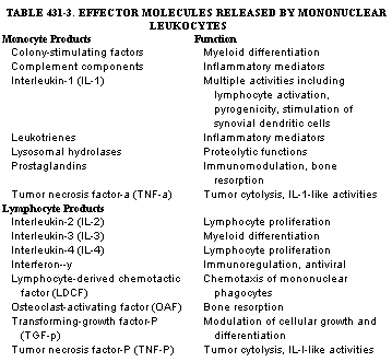
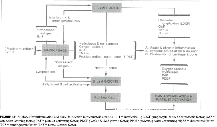
FIGURE 431-3. Requirements for phagocytosis. Particulate antigen binding to the membrane of phagocytic cells initiates cellular responses that lead to envelopment of the antigen. The process is enhanced when the antigens have bound opsonins. TYPE II REACTIONS: TISSUE DESTRUCTION MEDIATED BY CYTOTOXIC ANTIBODY. The development of antibody to antigens on the surface of a host's own cells can lead to tissue destruction. Injury results from the binding of C-fixing antibodies to host tissue cells. Activation of the C cascade leads to the release of inflammatory mediators and the accumulation of inflammatory cells. Release of lysosomal enzymes and toxic oxygen radicals by inflammatory cells and direct cvtolvsis of tarizet cells through C action contribute to tissue destruction. A human disease resulting from antibody directed toward self tissues is Goodpasture's syndrome (see Ch. 63). The result of antibody deposition on pulmonary and glomerular basement membranes is the explosive onset of hemorrhagic pneumonitis and rapidly progressive glomerulonephritis. Spontaneous development of antibody to tissues characterizes certain rheurnatologic disorders, particularly systemic lupus erythematosus (SLE). Autoimmune hemolytic anemia occurs following the deposition of C-fixing antibodies plus C3 cleavage products on circulating red blood cells. As a consequence, the cells are rapidly destroyed either by macrophages in the reticuloendothelial system (particularly in the spleen) or, less commonly, by intravascular hemolysis mediated by C. Also common in SLE is idiopathic thrombocytopenic purpura, in which antibody develops against platelet antigen and leads to thrombocytopenia TYPE III REACTIONS: INFLAMMATION INITIATED BY IMMUNE COMPLEXES. The formation or deposition of certain types of immune complexes in local tissues produces an inflammatory response characterized by the accumulation of polymorphonuclear leukocytes within hours, followed by the influx of macrophages. Several mechanisms exist by which immune complexes initiate this reaction. The combination of IgM or IgG (subclasses 1, 2, or 3) antibodies with antigen leads to binding and activation of the first component of C. As a result, C4 and C2 are cleaved and activated, and then C3 is cleaved into two fragments. The larger fragment (C3b) binds to the immune complex; the smaller fragment (C3a) diffuses away. C3a. enhances vascular permeability, contracts venular smooth muscle, and degranulates mast cells and basophils. Cleavage of C5 releases the potent inflammatory polypepticle C5a. Diffusion of C5a from the site of immunologic reactions establishes a gradient of this chemoattractant. Polymorphonuclear leukocytes and macrophages detect this gradient and migrate to the site of immune complex deposition. Upon arrival, Fc, C3b, and iC3b receptors enhance complex binding to the phagocytic cells, and phagocytosis ensues (Fig. 431-3). If the amount of immune complex deposited locally is not great, the material can be phagocytized and digested without tissue destruction. If the amount is large or if a significant portion is lodged in vessel walls, permanent tissue destruction can ensue. Polymorphonuclear Ieukocytes and macrophages contain abundant lysosomal enzymes and are capable of producing toxic oxygen radicals. In the process of phagocytizing immune complexes, particularly when these complexes are not easily internalized, the cells release their lysosomal enzymes and oxygen radicals externally. The lysosomal enzymes are capable of cleaving additional C5, thereby producing more C5a. These processes, when occurring within vessel walls, produce vasculitis and can lead to hemorrhagic necrosis and local tissue destruction. Tissue damage initiated by C-fixing immune complexes can occur following the formation of antigen-antibody complexes in local tissue sites (an Arthus-type reaction) or in the circulation (a serum sickness reaction). Serum sickness reactions occur when an individual develops C-fixing antibody to a circulating antigen. As antibody is produced, antigen-antibody complexes form in the circulation. During the early phase of antibody synthesis, the amount of antibody available for binding is small so that the complexes are formed in a setting of great antigen excess. Such complexes are not pathogenic. As antibody production increases, usually by seven days after antigenic exposure, the immune complexes become larger and the ratio of antigen to antibody decreases. When the complexes are in slight antigen excess, they tend to be deposited in the walls of small blood vessels, where they initiate inflammatory lesions. Palpable purpuric skin lesions (leukocytoclastic vasculitis), arthritis, glomerulitis, and fever, as well as depressed serum C levels, are common clinical manifestations. As antibody production continues, the remaining immune complexes in the circulation increase in size and are rapidly cleared by the reticuloendothelial organs. If the exposure to antigen ceases, the illness resolves. Examples of both types of inflammation initiated by immune complexes are common in rheumatic diseases such as rheumatoid arthritis and SLE. Certain animals develop spontaneous immune complex diseases that bear striking similarities to human SLE. The F, hybrid cross between New Zealand Black (NZB) and New Zealand White (NZW) mice (NZB/W FJ develops an immunologic disorder characterized by circulating antibody to nuclear proteins, glomerulonephritis, autoimmune hemolytic anemia, and vasculitis. In contrast to the NZB/W F, hybrids, NZB mice develop severe autoimmune hemolytic anemia but insignificant glomerulonephritis. NZW mice do not develop spontaneous autoimmune disease. NZB/W disease illustrates the contribution of genetic, immunologic, and infectious factors to the production of a spontaneous immune complex disease. Genetically, multiple autosomal genes appear to be involved. Immunologically, the animals have heightened B-cell activity and abnormalities of regulatory T-cell function, including depressed suppressor function. A direct role of helper T cells in stimulating autoantibody responses has also been inferred, since therapy of mice with monoclonal antibodies to helper cells ameliorates disease. The disease itself appears to occur when the NZB/W mice produce unusually large amounts of antibody to Gross leukemia virus, an agent that infects many normal mouse strains but usually produces no disease. In the NZB/W mice, however, antigen-antibody complexes develop and deposit in a granular "lumpy-bumpy" immunofluorescent pattern in the renal glomerulus, leading to immune complex-induced glomerulonephritis. The circulating immune complexes cause a fall in the serum C titer, and a systemic vasculitis occurs. Unique subpopulations of anti-DNA antibodies have also been implicated in renal immune complex deposition in these animals. Recently, mice with genetic backgrounds quite different from those of NZB/W have also been shown to develop spontaneous immune complex disease. MRL-Ipr/lpr and BXSB animals develop an illness characterized by circulating immune complexes, depressed serum C, and immune complex nephritis. Single gene abnormalities appear to be responsible for the autoimmune disease in mice of these strains. In MRL mice, an autosomal recessive gene termed 1pr leads to autoantibody production, while in BXSB mice a Y-chromosome linked gene termed Y-, for autoimmune accelerator, plays a similar pathogenetic role. As in the NZB/W mice, one of the antigens appears to be an oncornavirus, protein. Mice of the MRL strain differ from the NZB/W F, in that they produce antibody to a nuclear antigen termed Sm. This type of antibody has been previously found only in humans with SLE. The immune defect in MRL mice is associated with increased helper T cell activity and elaboration of a T cell factor that stimulates B cells. MRL-Ipr/lpr mice also develop an erosive arthritis with similarities to rheumatoid arthritis. Prior to cartilage and bone destruction, dedifferentiated synovial cells migrate across cartilaginous surfaces and ultimately invade the hard tissues of the joint. The requirement for a genetic predisposition along with exposure to the appropriate infectious agent or other environmental factor is also likely in human SLE and rheumatoid arthritis. Humans with SLE usually share similar histocompatibility antigens at the DR locus. Moreover, abnormal T cell suppressor function is seen not only in patients but also in family members. Other human illnesses that appear to occur secondary to circulating or localized immune complexes include certain adverse reactions to drugs, hypersensitivity pneumonitis, and reactions to viruses such as hepatitis B virus. TYPE IV REACTIONS: INFLAMMATORY REACTIONS INITIATED BY MONONUCLEAR LEUKOCYTES. Lymphocyte-initiated inflammatory reactions are termed "delayed hypersensitivity" because maximal inflammatory cell accumulation does not appear for 48 to 72 hours after secondary antigenic exposure. For example, if an individual previously sensitized to the tubercle bacillus is injected locally with antigen from this organism, a delayed type of inflammatory response ensues. The foreign material is encountered first by macrophages and dendritic cells, which partially digest the antigen. This altered form of antigen is recognized by small lymphocytes that contain specific surface receptors for the antigen. Exposure to the antigen initiates synthesis and release of cytokines (Table 431-3), which diffuse to areas of the vessel wall closest to the immunologic event. Increased vascular permeability ensues; chernotactic gradients that attract macrophages and other lymphocytes result in inflammatory cell accumulation. Granulocytes precede the mononuclear cell influx, but their number is far less than in the inflammatory response mediated by immune complexes. Lymphokines also activate the macrophages, which become more metabolically active, develop higher levels of hydrolytic enzymes, and are better able to bind to and destroy tumor cells or many intracellular parasites. Lymphocytes at the inflammatory site release lymphokines that recruit other nonsensitized lymphocytes, thus expanding the clones of cells capable of recognizing and responding to the specific antigen. If successful in complete destruction of the antigen, the inflammatory response resolves and produces no tissue necrosis. However, if the antigen is large in quantity or is difficult to digest, the inflammatory process continues. New cells arrive to replace the dying cells already present, resulting in the release of proteolytic enzymes and toxic oxygen radicals. Lesions typical of delayed hypersensitivity reactions are seen in mycobacterial and fungal diseases, sarcoidosis, and a number of rheumatologic disorders, including polymyositis and the granulomatous vasculitides. MECHANISM OF TISSUE DESTRUCTION IN RHEUMATOID ARTHRITIS The characteristic tissue reaction in rheumatoid arthritis is synovitis, in which the normally thin, loose connective tissue is replaced by a rich infiltrate of lymphocytes, macrophages, and plasma cells (see Fig. 431-IB). In the synovial fluid, the predominant inflammatory cells are the polymorphonuclear leukocytes. The cells are metabolically active: The plasma cells produce rheumatoid factors, and the mononuclear cells produce cytokines. Rheumatoid factors are immunoglobulins of the IgM, IgG, or, rarely, IgA class. These factors bind to the Fc portion of IgG antibodies, which either have combined with antigen ' or have been aggregated or denatured. Polymorphonuclear leukocytes taken from synovial fluids contain inclusions of immune complexes, many of which contain rheumatoid factors. Synovial fluid C levels are depressed in relation to serum levels. The turnover of C components, particularly of the classic pathway, is markedly enhanced in rheumatoid synovial fluid. Cleavage products such as C5a are present in rheumatoid synovial fluid, as are hydrolytic enzymes derived from inflammatory cells, kinins, LTB, and the inflammatory neuropeptide, substance P. The sequence of events leading to synovitis and destruction of surrounding structures in rheumatoid arthritis can be envisioned as follows: Some as yet undefined antigen localizes in the synovium and is phagocytized by Type A synovial cells. The antigen is not completely destroyed and diffuses into the joint space, where it may adhere to cartilage. Binding of the processed antigen to B lymphocytes induces their differentiation to plasma cells, which then produce antibodies including rheumatoid factors upon chronic stimulation. Activation of T lymphocytes triggers lymphokine synthesis followed by blastogenesis. The role of viruses as stimulators of the immune response in rheumatoid arthritis must be considered. Rheumatoid synovial explant cells established in permanent lines exhibit many characteristics of virally transformed lymphocytes. Cell lines of the B lymphocyte variety frequently contain antigens of the Epstein-Barr virus (EBV). Sera from approximately 65 per cent of patients with rheumatoid arthritis contain antibody, called a rheumatoid arthritis precipitin (RAP), to nuclear antigens present in human lymphoblastoid cell lines infected with EBV. The antigens have been termed rheumatoid arthritis nuclear antigens (RANAs). RAPs are commonly present in sera of patients who are rheumatoid factor positive but are also found in the sera of patients who are rheumatoid factor negative. The specificity of RAPs for rheumatoid arthritis has been questioned, as RAPs can be present in up to 20 per cent of normal individuals. In rheumatoid arthritis, there is no firm evidence to relate EBV causally to the disease, and serum antibody levels to EBV are not increased in the sera of rheumatoid arthritis patients. However, EBV does act as a polyclonal stimulator of antibody production and increases the mitogenic activity of lymphocytes. Another potential viral etiology may be related to parvovirus infection. This virus has been isolated from rheumatoid synovia, and acute parvovirus infection can cause at least a transient polyarthritis. Regardless of the initiating agent, combination of antibody with antigen, as well as combination of antigen-antibody complexes with rheumatoid factors, or self-association of rheumatoid factors, activates C as well as the kinin-forming system. This results in the production of inflammatory products such as C5a, arachidonic acid metabolites, kinins, and fibrinopeptides, which diffuse into the synovial fluid and to synovial blood vessels. These phlogistic agents enhance vascular permeability and attract polymorphonuclear leukocytes and macrophages. Polymorphonuclear leukocytes ingest the abundant immune complexes in the fluid, release lysosomal enzymes, and generate superoxide anions. This causes destruction of hyaluronate polymers in the joint fluid, as well as injury to cartilage. Cytokine and growth factor (i.e., interleukins, TNF, TGF-P, and platelet derived growth factor [PDGFJ) production in the synovium leads to the further accumulation and activation of macrophages, fibroblasts, and lymphocytes. The unique structure of the joint space is important, as enzymes present in synovial fluid or released and synthesized locally by the cells of the proliferative synovial lesion contribute to the pathology. The cartilage-degrading lysosomal enzymes collagenase and elastase are primarily derived from inflammatory cells. Proteinases released by dying cells may aid in superficial cartilage destruction by virtue of their role in uncrosslinking collagen fibrils, thus increasing their susceptibility to enzymatic degradation. Macrophages produce prostaglandins, hydrolytic enzymes, collagenase, plasminogen activator, IL-1, TNF-ot, and TGF-P. The most abundant source of collagenase in the rheumatoid synovium is a synovial cell that has an unusual dendritic appearance, expresses la antigen, and is adherent to glass but is nonphagocytic. Collagenase synthesis by this cell is greatly enhanced by IL1. With ongoing synovitis, early changes in cartilage involve the loss of proteoglycan content, often manifested microscopically as diminished metachromatic staining. In addition to collagenases, lysosomal proteinases can degrade aggregates of proteoglycans, and, once released from cartilage, these solubilized components are then sensitive to further enzymatic attack. The migration of the synovium (pannus) onto cartilage may be directed by the depositton of antigen there. In some synovial biopsies from patients with rheumatoid arthritis, tumor-like proliferations of dedifferentiated synovial cells can be found migrating across and into cartilage. The histopathology in these instances is similar to that seen in MRL mice. The final stage of the destructive process, demineralization of bone, may result from combined elements present initially in the inflammatory and later in the proliferative responses. Dernineralization must occur in bone before tissue is susceptible to collagenolytic enzymes. In rheumatoid synovitis, prostaglandins stimulate calcium release from bone matrix; other arachidonic acid metabolites are responsible for long-term leaching of mineral from bony matrix. In addition, bone dernineralization is enhanced by heparin, which is released upon the degranulation of mast cells. ' Cellular mechanisms may also be operative in dernineralization in that lymphocytes produce an osteoclast-activating factor (OAF). In addition, connective tissue activating peptides (CTAPs) and specific lymphokines such as lymphocyte-derived chernotactic factor for fibroblasts (LDCF-F) attract and stimulate fibroblasts to produce collagen and may contribute to the ultimate fibrosis in the destroyed ankylosed joint. The net effect, resulting from either persistence of antigen or disordered regulation of T and B cell activation, is a chronic inflammatory response in the synoviurn (Fig. 431-4). Continued cellular proliferation and influx lead to synovial proliferation and its invasion into surrounding structures. Diffusion of collagenase, PGEs, hydrolytic enzymes, and lymphokines into cartilage and bone results in erosion of these tissues. Rheumatoid arthritis thus illustrates the devastating local tissue destruction that results from chronic inflammatory reactions produced by the immune complex and delayed hypersensitivity types of immune responses. |
ENTRANCE | HOME | 1 | 2 | 3 | 4 | LINKS | FUN STUFF | BULLETIN BOARD | BOOK STORE | DISEASES | SEARCH