|
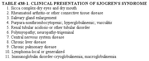
DEFINITION. Sjogren's syndrome is a chronic inflammatory and autoimmune disease in which the salivary and lacrimal glands undergo progressive destruction by lymphocytes and plasma cells, resulting in decreased production of saliva and tears. The term autoimmune exocrinopathy has been introduced. The spectrum of this illness includes a primary form (sicca complex), a secondary form accompanying rheumatoid arthritis (or occasionally another connective tissue disease), and a form characterized mainly by lymphoproliferation of either a benign infiltrative or a malignant nature. Females are involved 10 times more commonly than males. PATHOGENESIS. The several factors involved in the etiology of autoimmune diseases such as Sjogren's syndrome include genetic, immunologic, hormonal, and probably infectious (? viral). The discovery of the immune response (IR) genes, which exist in linkage disequilibrium with other genes in the major histocompatibility complex, has helped distinguish primary from secondary Sjogren's syndrome. The former is associated with HLA 138 DR3, whereas the latter is associated with DR4 (when rheumatoid arthritis is the accompanying illness). Recently, gene interaction between DQI and DQ2 has been associated with marked hypergammaglobulinemic and high-titered autoantibody responses. The presence of la molecule and Ebstein-Barr virus (EBV) in salivary gland epithelium has also been noted. The predominant female incidence of Sjogren's syndrome may relate to the ability of sex hormones to modulate the immune response, with estrogen contributing to immune hyperactivity. CLINICAL MANIFESTATIONS. The varied clinical presentations of Sjogren's syndrome are shown in Table 438-1. Ophthalmologic (Keratoconjunctivitis Sicca). The patient may notice accumulation of thick ropy secretions along the inner canthus owing to a decreased tear film and an abnormal mu us component. Related complaints include a characteristic foreign body sensation ("sandy or gritty eyes"), photosensitivity, eye fatigue, decreased visual acuity, and the sensation of a "film" across the field of vision. Desiccation can cause small superficial erosions of the corneal epithelium. Slit lamp examination may reveal filamentary keratitis (filaments of corneal epithelium and debris) in severe cases. Salivary. Complaints resulting from dryness of the mouth are varied. The "cracker sign" describes the difficulties encountered in trying to eat dry foods without sufficient lubrication. Many subjects require frequent ingestion of liquids. They may resort to carrying water bottles or candy in purse or pocket. Additional features include oral soreness, adherence of food to buccal surfaces, fissuring of the tongue, and dysphagia. Angular cheilitis resulting from superimposed candidiasis may occur. Patients may lose the ability to discriminate foods on the basis of taste and smell. Dental caries are accelerated. The parotid gland enlarges in many patients secondary to cellular infiltration and ductal obstruction. Usually asymptomatic and self-limited, the enlargement can be recurrent and associated with pain and erythema. Focal infiltrates of lymphocytes are also found in the minor salivary glands of the lower lip. When biopsied, these lesions provide histologic confirmation and quantification of the degree of infiltration.
Other Symptoms. Dryness may also involve the nasal mucosa, leading to recurrent epistaxis, and may extend throughout the upper respiratory tract, causing hoarseness, recurrent bronchitis, and pneumonitis. Eustachian tube blockage can result in conduction deafness and chronic otitis. Dysphagia may be ascribed to several causes: decreased saliva, infiltration of the glands of the esophageal mucosa, esophageal webbing, and abnormal motility. Other exocrine gland functions may be affected, leading to loss of pancreatic secretions, hypo- or achlorhydria, dermal dryness, and lack of vaginal secretions. Extraglandular Involvement. Extraglandular involvement occurs more frequently in patients with primary than secondary Sjogren's syndrome. Dependent nonthrombocytopenic purpura is generally associated with hyperglobulinemia. Raynaud's phenomenon is present in 20 per cent of patients. A diffuse interstitial pneumonitis resulting from lymphocytic infiltration may cause dyspnea. Obstructive disease (in the absence of smoking) may result from lymphocytic infiltration surrounding small airways. The most common renal abnormalities involve the tubules, particularly overt or latent renal tubular acidosis and hyposthenuria. The presence of glomerulonephritis should suggest coexisting systemic lupus erythematosus (SLE), cryoglobulinemia, or immune complex deposition. Peripheral and cranial neuropathy have been associated with vasculitis involving the vasa nervorum. A variety of CNS manifestations has recently been described. Lymphoproliferation and Lymphoma. The incidence of lymphoma is increased 44-fold in Sjogren's syndrome. Pseudomalignant or malignant lymphoproliferation may be present initially or may develop later in the illness. Most lymphomas belong to the B cell lineage, as demonstrated by immunophenotyping and immunogenotyping. Monoclonal immunoglobulin B cell proliferations in Sjogren's syndrome patients include Waldenstr6m's macroglobulinemia, light chain myeloma, and non-IgM monoclonal gammopathies (IgG K and IgA X). A diminution of a previously elevated Ig class may signify malignant transformation. Pseudolymphoma is an intermediate stage in this transition from benign to malignant lymphoproliferation. Other clinical indications of an increased risk of malignancy include persistent or greatly increased parotid swelling, generalized lymphadenopathy, and splenomegaly. DIAGNOSIS. Clinical. The presence of dry eyes is suggested by a positive Schirmer test (less than 5 mm of wetting per five minutes, unanesthetized), but the frequency of both false-negative and false-positive results is high. The pattern and intensity of staining with rose bengal dye and slit lamp examination are more reliable in diagnosis. The presence of filamentary keratitis and corneal ulcerations indicates advanced keratoconjunctivitis sicca. Diminution in stimulated parotid flow rate (PFR) (<5 ml per gland in 10 minutes) is a sensitive indicator of xerostomia. Salivary scintigraphy, which measures the uptake, concentration, and excretion of 11-Tc-pertechnetate by the major salivary glands, is a sensitive index of glandular function. Scintigraphy is expensive, however, and offers no advantage in diagnostic sensitivity over minor salivary gland biopsy. Lip biopsy is a sensitive and specific diagnostic procedure, is well tolerated by the patient, and causes no disfigurement. In addition to confirming the diagnosis, biopsy allows quantification of the degree of lymphocytic infiltration and tissue damage. Aggregates of lymphocytes within the acinar tissue are scored, each aggregate of 50 or more cells representing a focus. The number of foci within 4 sq mm of glandular tissue is determined and constitutes the focus score. A focus score of more than 1 is characteristic of Sjogren's syndrome and is seen in less than 1 per cent of both normal and autopsy controls. The diagnosis of Sjogren's syndrome is based upon the presence of two of the following three criteria: (1) focus score of more than I in the labial salivary gland biopsy, (2) keratoconjunctivitis sicca, and (3) an associated connective tissue or lymphoproliferative disorder. Clinically, a "sicca-like" syndrome may be caused by a number of other disease processes, including hyperlipoproteinemias IV and V, hemochromatosis, sarcoiclosis, and amyloidosis. Use of anticholinergic drugs as well as a number of other medications may be the single most frequent cause of xerostomia. Thus, it is essential to establish the presence of focal lymphoid infiltrates and autoimmunity in a patient suspected of having Sjogren's syndrome. Laboratory. Autoantibodies are common in Sjogren's syndrome. Rheumatoid factor may be found in 75 to 90 per cent; antinuclear antibodies may be positive in 50 to 80 per cent. Multiple organ-specific antibodies are noted, including antibodies directed against gastric parietal, thyroid microsomal, thyroglobulin, mitochondrial, smooth muscle, and salivary duct antigens. An autoantibody to a nucleoprotein antigen called SS-13 (also termed La) occurs in up to 90 per cent of patients with primary Sjogren's syndrome and to a lesser extent in Sjogren's syndrome accompanied by SLE. Antibodies to a related nucleoprotein SS-A (also termed Ro) are less specific for Sjogren's syndrome, also occur in SLE, and are associated with vasculitis. An antibody (RAP) to an EBV-related nuclear antigen (RANA) occurs in secondary Sjogren's syndrome with RA. Persons with Sjogren's syndrome manifest B cell hyperactivity. Evidence for this includes the polyclonal hyperglobulinernia seen in over 50 per cent of patients and the presence of numerous autoantibodies and circulating immune complexes. The lymphoid infiltrates in the salivary glands synthesize immunoglobulins locally. Serum hyperviscosity may result from either macroglobulinernia or polymerizing IgG with rheumatoid factor activity which forms intermediate complexes. Cryoglobulinernia may be present, as well as vasculitis and glomerulonephritis. A high proportion of patients with Sjogren's syndrome have circulating immune complexes as measured by C1q binding and Raji cell assays. Serum levels of complement are only infrequently low. Peripheral blood T lymphocytes are decreased in about one third of patients. Immunoglobulin-positive lymphocytes in peripheral blood may be increased slightly. Abnormalities in T cell function may be present, particularly in patients with lymphoproliferative or other systemic features. These patients tend to have alterations in T cell subsets and decreased autologous mixed lymphocyte responses. Natural killer (NK) cell activity is also diminished and may contribute to the emergence of lymphoid malignancy. TREATMENT. Treatment of Sjogren's syndrome is aimed at symptomatic relief and limiting the damaging local effects of chronic xerophthalmia and xerostomia. Ocular dryness responds to the use of artificial tears, which can be applied every one to three hours. A slow-release tear is also available. Soft contact lenses may be used to protect the cornea but increase the risk of infection. Saran Wrap occlusion or diving goggles may be worn at night in an attempt to prevent tear evaporation. Topical steroid use should be avoided unless specifically indicated, because corneal thinning and subsequent perforation may occur. The use of diuretics, many antihypertensive drugs, and antidepressants may further diminish lacrimal and salivary gland function. Xerostomia is difficult to treat but may be relieved with water, chewing gum, or sugar-free candies given as sialagogues. Scrupulous care of teeth is imperative; patients should avoid a high sucrose intake or the frequent use of sugar-containing candies to decrease oral dryness. Vigorous dental plaque control and topical application of fluoride should be used regularly. Oral candidiasis may be treated with Mycostatin tablets for a prolonged course, with separate treatment of dentures. Vaginal dryness can be treated with propionic acid gels. Only those patients with severe functional disability or lifethreatening complications warrant corticosteroid or immunosuppressive therapy. Prednisone may suppress parotid swelling and improve the restrictive component of pulmonary disease. Cyclophosphamide has decreased extraglandular lymphoid infiltrates and improved exocrine gland function in some individuals. Its use has been restricted to those patients with severe renal, skin, or pulmonary manifestations. |
ENTRANCE | HOME | 1 | 2 | 3 | 4 | LINKS | FUN STUFF | BULLETIN BOARD | BOOK STORE | DISEASES | SEARCH
