ENTRANCE | HOME | 1 | 2 | 3 | 4 | LINKS | FUN STUFF | BULLETIN BOARD | BOOK STORE | DISEASES | SEARCH
Barton F. Haynes
|
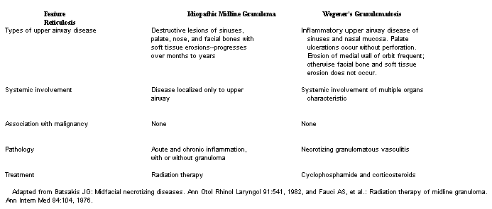
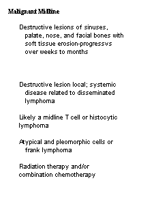
WEGENER'S GRANULOMATOSIS DEFINITION. Wegener's granulomatosis is a distinct clinical form of systemic necrotizing vasculitis consisting of (a) necrotizing granulornatous vasculitis of the upper and lower respiratory tracts, (b) focal necrotizing glomerulonephritis, and (c) systemic small vessel vasculitis involving numerous organ systems. ETIOLOGY. The cause of the disease is unknown. It is thought to be a hypersensitivity reaction to unknown inhaled antigen(s). No familial, geographic, or occupational exposure factors have been associated with the disease. The incidence of HLA B8 antigen may be increased in patients with the disease. INCIDENCE AND PREVALENCE. Wegener's granulomatosis is an uncommon but not rare disease that can affect any age-group. The mean age at onset is 40 years, with a male:female ratio of 3:2. PATHOLOGY AND PATHOGENESIS. The typical histopathologic lesion is necrotizing vasculitis of small arteries and veins, usually with granuloma formation in the surrounding cellular infiltrates Biopsy of paranasal sinus, nasopharyngeal, or tracheal lesions may show acute or chronic inflammation or frank vasculitis with or without granulomas. Pansinusitis, nasal crusting with drainage, and serous otitis may result, as well as nasal septal perforation and saddle nose deformity. Pulmonary lesions are present in 95 per cent of patients, with granulomas and vasculitis the common findings in biopsy material. Lung infiltrates are typically multiple, nodular, bilateral lesions that frequently cavitate. Less frequently, obstructive endobronchial lesions that lead to airway obstruction and atelectasis, or pleural lesions leading to pleural effusions, are found. Renal involvement is due to focal segmental glomerulonephritis that can lead to glomerular necrosis, crescent formation, and rapidly progressive renal failure. Any other organ system, most commonly skin and eyes, can be involved, with small vessel vasculitis with or without granuloma formation. Although the specific mechanisms that lead to granulomatous vasculitic lesions in Wegener's granulornatosis are poorly understood, there is considerable evidence suggesting that disordered immunity with both antibody and cell-mediated tissue damage occurs. Approximately 50 per cent of patients have elevated levels of circulating immune complexes, and positive rheumatoid factor and hypergammaglobulinemia with elevations of serum IgA and IgG are common. Deposition of IgG and complement components as well as fibrin can be found in some renal biopsies. Pulmonary infiltrates show redominantly T cells and macro phages in thepnulomatous feZons as weff as pofymorphonuclear cells (PMN) in and around inflamed vessels. Some studies have suggested an abnormality of PMN's in this disease with chernotactic defects, antineutrophil antibodies, and intravascular lysis of leukocytes. It is likely that there are more than one immunologic mechanism occurring, such that an abnormal or exaggerated ~ntibody response to an inhaled antigen could lead to an immune-complex triggered macrophage-T cell granulomatous response centered in and around vessels. CLINICAL MANIFESTATIONS. The most common presentation of Wegener's granulornatosis is with upper and lower airway disease (90 to 95 per cent). Multiple organ systems may be involved during the course of the disease, including kidneys, joints, ears, and eyes. Common signs and ~ymptoms include purulent nasal discharge, fever, cough, nemoptysis, paranasal sinus pain, nasal mucosal ulceration, and saddle nose deformity. Lung involvement can be asymptomatic, with nodular infiltrates seen on routine radiograph. Renal disease is seen in 85 per cent of patients and is a critical determinant of the clinical outcome. Renal manifestations include hematuria, azotemia, proteinuria, and pedal edema. Renal disease can be smoldering, but more often when untreated rapidly progresses to irreversible renal failure. Nearly 70 per cent of patients have some form of joint involvement during the course of the disease. Of these, one third have a nondeforming arthritis, usually in ankles and knees. The remaining two thirds have symmetric polyarticular arthralgias. Eye involvement occurs in 60 per cent of patients and is manifested as proptosis, scleritis, conjunctivitis, uveitis, dacryocystitis, and retinal or optic nerve vasculitis. Corneoscleral ring ulcers may progress to scleral perforation. Proptosis is usually due to contiguous sinus involvement with extension of granulomatous inflammation into the orbit. Nervous system disease occurs in 20 per cent of patients, with mononeuritis multiplex the most common manifestation. Central nervous system involvement can be in the form of cranial nerve dysfunction, diffuse cerebral vasculitis, or hypothalamic granulomas with diabetes insipidus. Heart involvement is manifested by pericarditis or coronary vasculitis. Less common manifestations include thyroiditis, mastoiditis, parotid masses, nasolacrimal duct obstruction, ear pinna and tympanic membrane granulomas, ulcerating breast masses, and anosmia. Characteristic laboratory abnormalities include elevated erythrocyte sedimentation rate, neutrophilic leukocytosis, anemia, and positive test for serum rheumatoid factor. Serum antineutrophil antibodies against a cytoplasmic antigen have been reported to be useful for diagnosis and monitoring disease activity. DIAGNOSIS. 'The diagnosis should be strongly considered when findings of upper or lower respiratory tract disease, renal disease, and vasculitis involving other organ systems are present. The disease should also be considered when any of the typical disease manifestations occur as an isolated finding (such as proptosis due to granulomatous inflammation and vasculitis) in an otherwise well patient. To establish a definitive diagnosis, a patient should have evidence of clinical disease in at least two of the following three areas: upper airways, lung, and kidney. Biopsy results should show disease in at least one and preferably two of these organ systems, with lung tissue providing the source of highest diagnostic yield. Open lung biopsy is the procedure of choice to obtain adequate tissue for diagnosis. Percutaneous renal biopsy is important for both diagnosis and documentation of extent of renal disease. Differential diagnosis should include other diseases that can cause pulmonary-renal syndromes such as Goodpasture's syndrome (see Ch. 62 and 81). Idiopathic midline granuloma (see below) destroys facial and palate bones and cartilage but is not a systemic vasculitis and does not involve lungs or kidneys. Lesions of Wegener's granulornatosis do not perforate the palate or erode through major bony structures of the face and upper airway, although they may destroy the medial wall of the orbit, called the lamina papyracea. Another disease frequently confused with Wegener's granulomatosis is lymphomatoid granulomatosis. This disease is characterized by infiltration of various organs with a polymorphic cellular infiltrate consisting of atypical lymphoid and plasmacytoid cells together with granulomatous inflammation in an angiocentric pattern. The disease primarily involves lungs, skin, kidneys, and central nervous system, but not upper airways. Renal involvement is not a glomerulonephritis, but rather is due to interstitial infiltration with masses of atypical lymphoid cells. Lymphomatoid granulomatosis, unlike Wegener's granulornatosis, is not an inflammatory vasculitis, but more likely represents invasion of vessels with premalignant T cells. Up to one half of cases evolve into a frank T cell lymphoma that responds poorly even to combined chemotherapy regimens for non-Hodgkin's lymphoma. TREATMENT AND PROGNOSIS. The treatment of choice for Wegener's granulomatosis is a combination of corticosteroids and a cytotoxic agent, of which the most efficacious is cyclophosphamide. Irreversible organ system dysfunction can occur if conservative therapy (such as with corticosteroids alone) is attempted. In patients with active but stable multisystem disease, oral cyclophosphamide should be given in a dose of 1 to 2 mg per kilogram per day, with the dose adjusted to maintain the total leukocyte count above 3000 per cu mm and the PMN count above 1000 to 1500 per cu mm. Weekly monitoring of the white blood cell count is essential to avoid severe leukopenia and infectious complications. In patients with fulminant disease, such as CNS vasculitis, severe pulmonary involvement with hypoxemia, rapidly progressive peripheral neuropathy, or rapidly progressive renal failure, cyclophosphamide may be given intravenously in a dose of 4 mg per kilogram per day for three days, with change to the lower oral dose regimen thereafter. Patients should be treated for one year after remission has been achieved, then cytotoxic drug therapy stopped and the patient re-evaluated periodically for disease relapse. Because of the risk of serious complications with cytotoxic therapy (see Ch. 176) and frequent symptomatic infection of damaged respiratory tracts with microorganisms suchas Staphylococcus aureus, the presence of persistent vasculitis should be documented prior to continuation of long-term cytotoxic therapy (greater than one year) for presumed active disease. Corticosteroids should be administered in an oral daily dose regimen (usually prednisone I mg per kilogram per day) or in divided doses (every six hours) for fulminant cases for an initial one to two weeks, followed by tapering to an alternateday regimen (see Ch. 30). After three to six months, corticosteroids can usually be discontinued altogether. With this regimen remissions can be obtained in 90 per cent of patients with Wegener's granulomatosis. Those patients who have gone on to end-stage renal failure have had successful renal transplants while on this regimen. Patients who relapse while on little or no medication require diagnosis of the cause of relapse. In a previously treated patient with a new pulmonary infiltrate, it is critical to distinguish between a lung infection and a recurrence of pulmonary vasculitis. MIDLINE GRANULOMA DEFINITION. Idiopathic midline granuloma, also known as idiopathic midline destructive disease, is a progressive, localized process that predominantly involves the nose, paranasal sinuses, and palate, with erosion through bone and soft tissues, frequently involving the face. ETIOLOGY. The cause of idiopathic midline granuloma is unknown. No primary infectious or neoplastic cause has been found. An exaggerated hypersensitivity reaction to unknown inhaled antigens has been postulated. PATHOLOGY. Biopsy demonstrates necrosis and acute and chronic inflammation. Mucosal surfaces of the nose and paranasal sinuses are frequently ulcerated, and inflammation invades and destroys adjacent cartilage and bone. The cellular infiltrate comprises polymorphonuclear leukocytes, lymphocytes, macrophages, plasma cells, and less frequently multinucleated giant cells with well-formed epithelial granulomas. Although perivascular infiltrations and vessel destruction due to widespread inflammation are common, a primary vasculitis is generally not seen. Foci of atypical lymphocytes or histiocytes suggest an underlying lymphoma or other midline neoplasm, rather than midline granuloma. CLINICAL MANIFESTATIONS. Most patients with idiopathic midline granuloma have active sinusitis with superimposed bacterial infections. Symptoms begin with nasal stuffiness and crusting and progress to purulent nasal discharge and bleeding. Ulcerations may appear on the nasal septum, palate, or nose. Perforation and destruction of the nasal septum can occur, resulting in a saddle nose deformity. Destruction of the soft and hard palate permits reflux of food into the upper airway during eating. Paranasal inflammation with swelling frequently results in nasolacrimal duct obstruction and dacryocystitis. Relentless midline inflammation with necrosis can result in widespread, mutilating destruction of facial bones and tissues, with erosion into the central nervous system leading to meningitis, or into major blood vessels leading to life-threatening hemorrhage. Erosion can cause retro-orbital masses, orbit destruction, and blindness. Loss of smell is common, and recurrent sinus infection with Staphylococcus aureus is routine. Most patients are free of systemic signs and symptoms except when superimposed bacterial infections occur. There are no characteristic laboratory findings. Computerized tomograms and radiographs of the mid line structures often show dramatic destruction of facial bones with evidence of widespread sinus inflammation. DIAGNOSIS. The diagnosis is made from the characteristic clinical presentation of locally destructive lesions restricted to the upper respiratory tract and absence of histologic evidence of Wegener's granulornatosis or lymphoma. Although midline granuloma is a distinct disease, the diagnosis is made by excluding other diseases with similar findings. While a large number of infectious, connective tissue, and inflammatory diseases can cause ulcerations of the nose and midline structures, the three entities that most often comprise the differential diagnosis are those listed in Table 441-1. Midline granuloma is a locally destructive disease that is relentlessly progressive over months to years. It is not a primary vasculitis and thus is clearly distinct from Wegener's granulomatosis, in which the lesions are not progressive, erode only cartilage and thin facial bones, and frequently heal spontaneously. Malignant midline reticulosis is a pleomorphic lymphoma, frequently of the T cell type, that presents with extensive mucosal ulceration, tissue necrosis, bone destruction, and fistula formation in facial midline tissues. The tempo of the disease is rapid, and, on repeat biopsy, atypical lymphoid cells or frank lymphoma is found. The diagnosis is a difficult one, however, since the clinical picture may suggest an inflammatory process rather than a tumor, and on biopsy the malignant nature of the process may be obscured by an intense inflammatory infiltrate, presumably in reaction to tumor antigens. Rarely, other lymphomas, mycosis fungoides, and nonkeratinizing squamous cell carcinomas can produce midfacial destructive lesions. Other less destructive diseases in the differential diagnosis are sarcoidosis (Ch. 69), relapsing polychondritis (Ch. 445). and necrotizing sialometaplasia, a selfhealing process of six to eight weeks' duration which presents with large ulcerations of the hard palate and a necrotizing process in minor salivary glands. Infectious diseases that should be ruled out include chronic bacterial infections, tuberculosis, syphilis, rhinoscleroma (due to Klebsiella rhinoscleromatis), actinomycosis, leprosy, phycomycosis, blastomycosis, candidiasis, histoplasmosis, coccidioidomycosis, and leishmaniasis. TREATMENT AND PROGNOSIS. Radiation therapy, approximately 5000 rads, to the midfacial area is the treatment of choice. Prednisone and cytotoxic drug regimens have generally not been successful. Surgical procedures on involved tissues can accelerate the destructive process. However, following radiation therapy, surgical debridement and aggressive local care measures, such as routine upper airway irrigation and treatment of bacterial superinfections, can aid the healing process. Reconstructive plastic surgery and prosthesis placement for patients with extensive facial disfigurement are often of benefit. In long-term survivors, close monitoring for radiation-induced upper airway neoplasia is essential. |

|
Types of upper airway disease
Pathology
|
 |
ENTRANCE | HOME | 1 | 2 | 3 | 4 | LINKS | FUN STUFF | BULLETIN BOARD | BOOK STORE | DISEASES | SEARCH