An increasing number of specialized procedures are available for evaluation of patients with articular disease. Although many are useful, very few are diagnostic of a specific disorder. Laboratory data must be combined with clinical evaluation in order to arrive at a working diagnosis.
SYNOVIAL FLUID. In the patient with undiagnosed articular disease and an associated joint effusion, examination of the synovial fluid is mandatory. The preferred method of joint aspiration (most frequently the knee) is through the extensor surface, where major blood vessels and nerves are sparse and the synovial pouch is more superficial. If the synovial fluid sugar is to be measured, the patient should have fasted for six hours if possible. Careful sterile technique virtually precludes infection, the one very rare complication of arthrocentesis. The needle should be at least 19 gauge. After appropriate draping and intracutaneous instillation of 1 to 2 per cent procaine or Xylocaine, the n6edle is inserted through skin and subcutaneous tissue. It meets a small amount of resistance when it reaches the capsule and finally passes easily into the joint cavity. Although minimal amounts of fluid will suffice for basic studies, as a rule 6 to 10 ml will allow the necessary laboratory testing.
The synovial fluid is then ideally allocated as follows: (1) an aliquot (1 to 3 ml) in a sterile tube with heparin for bacteriologic studies (cultures and Gram stain) or direct plating of an aliquot on appropriate culture medium; (2) an aliquot (I ml) in a clean nonsterile tube with heparin-for routine cytology, white cell count, and differential (can also be used for mucin test and crystal examination); (3) an aliquot (2 to 3 ml) without anticoagulant in a clean nonsterile tube-for color, viscosity, clot turbidity, mucin clot test and crystals (as well as proteins, if indicated); and (4) an aliquot (2 ml) in a clean nonsterile tube with preservative-for glucose analysis with parallel determination of serum glucose.
Cultures must be obtained on all synovial fluids, since indolent bacterial infections can mimic or be superimposed on well-defined articular disease. Blood agar medium should be used; but if gonococcal infection is suspected, chocolate agar or its equivalent should be inoculated at the bedside. Appropriate cultures should be obtained if tuberculosis or fungal infection is suspected. A direct Gram stain should be performed on a concentrated specimen from the first tube, sigun for 20 minutes in a clinical centrifuge.
Normal
synovial fluid is a clear, pale yellow, viscous liquid that does not clot. It
was appropriately named by Paracelsus because of its viscosity and physical
resemblance to egg white. The few cells normally present (less than 200) are
mononuclear. Although serous cavity fluids are plasma ultrafiltrates, synovial
fluid is unique owing to the presence of hyaluronic acid (about 0.3 gram per
deciliter) produced by the synovial lining cells. This large asymmetric molecule
further influences the composition of synovial fluid, for because of its steric
structure, some solute passage through the water surrounding the molecules may
be hindered. Thus, according to the concept of excluded volume, the size and
shape of the molecule play a large role; i.e., large molecules such as fibrinogen
and macroglobulin would be excluded, and small molecules would more easily enter
the compartment. The state of hyaluronate (as roughly determined by the
mucin clot test) may be a primary determinant of the nature of the synovial
fluid in various pathologic conditions.
When
a synovial membrane is inflamed for any reason, the white cell count in the
synovial fluid increases. In a rough fashion one can classify this fluid into
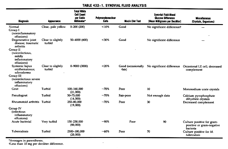
four groups (Table 432-1). Noninflammatory effusions (Group 1) occur when the
white cell count is normal or minimally increased, as in traumatic arthritis
or degenerative joint disease. Only rarely will such fluid have white cell counts
of over 2000 cells per cubic millimeter. Noninfectious mildly inflammatory effusions
(Group II) with white cell counts rarely over 5000 occur in systemic lupus erythernatosus
(SLE) and scleroderma. In noninfectious acute inflammatory effusions (Group
111) characteristic of classic rheumatoid arthritis, gout, pseudogout, and rheumatic
fever, the white cell count varies from 5,000 to 25,000 but may exceed 50,000
or even 100,000 cells per cubic millimeter. Finally, in inflammatory effusions
caused by infection (Group IV) the white cell count commonly varies from 25,000
to over 100,000 cells per cubic millimeter and in some instances resembles frank
pus. As the white cell count becomes elevated, the percentage of polymorphonuclear
leukocytes generally increases, the hyaluronate becomes degraded, and the synovial
fluid sugar falls.
Normal
synovial fluid does not clot owing to the absence of several clotting factors,
including fibrinogen. Pathologic fluids do contain clots, and their size is
roughly proportional to the degree of inflammation. Hyaluronate degradation
can be roughly assessed by diluting 1 ml of joint fluid with 4 ml of 2 per cent
acetic acid solution. When the mucin is normal, a tight ropy mass forms in a
clear solution. This is termed a "good" mucin. A softer mass with shreds is
a "fair" mucin, whereas a "poor" mucin shows shreds and soft small masses in
a turbid solution.
Under
normal conditions, the total synovial fluid protein is about one quarter that
of the blood. Multiple studies have been performed to assess whether specific
fractions might be related to specific disease states, but findings in general
are nonspecific. One may conclude that if the synovial fluid total protein is
over 2.5 grams per deciliter, the fluid is not normal, and that if it is over
4.5 grams per deciliter, there is significant inflammation.
Rheumatoid factors (antigamma globulins) are also found in the synovial fluid, occasionally when they are absent from the serum. Their presence and presumed local manufacture in the synovial membrane may be of diagnostic value. Antinuclear antibodies have been found not only in the synovial fluids of patients with SLE but also in those of patients with rheumatoid arthritis and several other connective tissue diseases. Since DNA, especially in the native form, can be found free in synovial fluids from individuals with a variety of disorders, it is probable that its presence may reflect nonspecific tissue damage.
Complement
levels in the synovial fluid depend upon rates of synthesis, catabolism, and
local consumption and have little clinical value. The only tests widely available
are immunoassays for C3 and C4 and functional assays for total complement (CH50).
In rheumatoid arthritis the serum complement is not depressed, whereas in SLE
it is low. Cryoproteins, predominantly of fibrinogen but including cryoglobulins
with DNA and IgG, are also found in synovial fluids of patients with rheumatoid
arthritis and other types of synovitis, and rheumatoid fluids may contain complexes
with Igs, rheumatoid factors, complement, and DNA.
The
intense inflammatory activity of various synovial diseases is associated with
the presence of a variety of enzymes in the synovial fluid. Some, but not all,
investigators have implicated lysosomal enzymes (e.g., myeloperoxidase, lactoferrin,
lysozyme, chymotryptic cationic protein) in the process of joint destruction.
Collagenase, collagen, and antibodies to collagen have also been extensively
studied. Such investigations have advanced our concepts of the pathogenesis
of synovitis (especially rheumatoid), but generally measurements of these substances
have not reached the routine clinical domain. Other proteins and peptides (C-reactive
protein, plasminogen, fibronectin, vasoactive peptides, and oxygenderived free
radicals) have also been measured in synovial fluid, but their precise role
in diagnosis is not clear.

Synovial
fluid normally contains little lipid, but it demonstrates increased lipid content
in rheumatoid effusions. Prostaglandin E, appears to be elevated in the fluid
of patients with inflammatory synovitis. The glucose content of synovial fluid,
which normally approximates that of serum, is markedly diminished in the presence
of infection and may show a modest decrease (10 to 30 mg per deciliter) in other
inflammatory effusions. Several of these nonspecific parameters are collectively
useful in evaluating the degree and type of inflammatory synovitis (Table 432-1).
Several types of crystals have been found in synovial fluids (Table 432-2). The two most important are monosodiurn urate, characteristic of gouty effusions, and calcium pyrophosphate dihydrate (CPPD), characteristic of the effusions of pseudogout (crystal deposition disease). Crystals that cause inflammation are usually 0.5 to 20 ~Lm in length, sparingly soluble in water, and capable of being phagocytized. At the peak of inflammation most are intracellular. Other crystals such as calcium hydroxyapatite, calcium oxalate, cholesterol, and corticosteroid esters may also be associated with inflammatory effusions.
Crystals are demonstrated in synovial fluid (collected without oxalate) by examining
a drop of synovial fluid placed on a slide and covered with a thin glass coverslip
for examination in the polarizing microscope. Birefringent materials demonstrate
two refractive indices when plane polarized light passes through them. The birefringence
is termed positive when the crystals (which then appear blue) are aligned parallel
to the slow rays of the retardation plate (first-order red plate compensator)
and negative when the crystals appear yellow when in parallel alignment (blue
when perpendicular). On polarization microscopy the monosodium urate crystals
demonstrate strong negative birefringence and are usually long (8 to 10 ILm)
and needle- like in appearance. They may occur extraellularly or within polymorphonuclear
or mononuclear cells. They are almost invariably in effusions associated with
acute gout and are virtually diagnostic. They may, however, be present in gouty
fluids between attacks. The CPPD crystals are often broader than urate crystals,
may show a faint "line" down their center, appear parallelopiped, and in the
polarizing microscope exhibit a weak positive birefringence. They have a significant
association with acute attacks of arthritis in patients with chondrocalcinosis
(articular cartilage calcification).
Other crystals identified in synovial fluid include hydroxyapatite (calcium phosphate) crystals, which are minute and identified only with special techniques; plate-like cholesterol crystals seen in some rheumatoid effusions; and the rare corticosteroid crystals found after intra-articular steroid treatment. All are capable of inducing a synovitis.
Under certain circumstances joint fluids, when nontraumatically aspirated, may demonstrate gross blood. One must consider bleeding disorders such as hemophilia, overdosage with anticoagulants, rare lesions such as pigmented villonodular synovitis and joint tumor, as well as neuropathic and traumatic forms of arthritis.
Careful examination of synovial fluid may on occasion reveal malignant cells, LE cells, sickled RBCs, fat droplets suggestive of fracture, or amyloid fragments.
SYNOVIAL MEMBRANE HISTOPATHOLOGY. Synovial membrane is a non-basement membrane-lined, highly vascular structure consisting of several cell types (mactophages, fibroblasts, and possibly intermediate cells). It proliferates extensively during inflammation, such that synovial biopsy is often useful in establishing a diagnosis. The procedure is simple and may be an extension of the synovial fluid aspiration technique, using a wider bore Parker-Pearson needle. A specimen can also be obtained by arthroscopy or open surgical biopsy.
Although in the common rheumatic diseases (rheumatoid irthritis degenerative joint disease, SLE) there are no comrnon pathognomonic synovial membrane findings, the patterns of histologic involvement may be diagnostically useful. For example, severe synovial lining proliferation and especially the presence of lymphoid follicles are characteristic of rheumatoid arthritis, whereas the SLE membrane shows only minimal hyperplasia but may show dense surface fibrin and perivascular mononuclear infiltrates; rarely a pathognomonic hernatoxylin body will be seen.
The procedure is very useful in the diagnosis of tuberculosis, atypical mycobacterial disease, or fungal disease; the demonstration of an organism in section (or in culture) or a caseating granuloma with giant cells virtually establishes the diagnosis. Other instances in which the synovial biopsy could be helpful include hemochromatosis (in which iron is seen in the synovial lining cell), pigmented villonodular synovitis (villous hypertrophy, hemosiderin deposits with numerous giant cells), tumors (malignant cells in synovium), and ochronosis (fragments of pigmented cartilage in synovium). Although in properly fixed tissue (use of absolute alcohol) one can observe the crystals associated with gout on histologic examination, the procedure is not necessary to establish this diagnosis, since examination of synovial fluid so commonly demonstrates the crystals.
ACUTE PHASE PHENOMENA. The ancient Greeks observed the sedimentation of blood following venesection and used it as a basic diagnostic tool. It was reintroduced by Fahraeus and refined by Westergren, whose method is in common use today. The sedimentation rate is only one, albeit the most popular, of the acute phase phenomena utilized by rheurnatologists to follow inflammation in their patients. Acute phase reactions refer to the increases in certain plasma proteins that occur after an extraordinary variety of tissue damage, i.e., toxic, chemical, infectious, inflammatory, or malignant. The functions of most of these proteins are not known, although presumably the damage led to increased protein synthesis via macrophage-derived mediators. Only the sedimentation rate and C- reactive protein (CRP) will be briefly discussed here.
The
basic measurement in the Westergren sedimentation rate is the rate of fall of
erythrocytes in plasma; when rouleaux formation occurs owing primarily to increases
in asymmetric proteins such as fibrinogen or macroglobulins (that alter the
red cell zeta potential), the red cells sediment more rapidly. The original
normal values are a 1 to 3 mm fall in one hour for men and a 4 to 7 mm fall
in one hour for women. These levels may be elevated during menses or by certain
drugs and appear to increase with aging. The cause of the last-named phenomenon
is not clear despite several reports that seem to document
it well. Many now accept sedimentation rates of 0 to 10 mm per hour
for men and 0 to 15 mm. per hour for women and suggest that the values
may be as much as 10 mm per hour higher after age 50. Whether these represent
changes in serum proteins with aging or undetected disease in the normal person
is not yet clear. In the rheumatic diseases, however, the value of this
determination is that when elevated (and it is often in the 30 to 100 mm per
hour range), it can be an index for following the severity of inflammation and,
when falling, for following the response to therapy. In addition, disorders
such as temporal arteritis are often associated with sedimentation rates of
over 100 mm per hour.
One must be constantly aware, however, of the lack. of specificity of this determination and that in selected series highly elevated sedimentation rates (over 100 mm per hour) are more commonly due to infections or malignancies. It is also documented that a small number of patients can have an active rheumatic disease (i.e., rheumatoid arthritis) with a normal sedimentation rate.
The CRP until recently has played little role in the evaluation of rheumatic diseases, although it has been the prototype acute phase reactarit-virtually absent in normal conditions and appearing in large quantities in inflammation. It was named for its property of precipitating with pneumococcal cell wall polysaccharide. In recent years it has been isolated and characterized as a pentameric structure, and its molecular weight and primary structure have been determined. It has many homologies with a protein (AP) found uniquely in amyloid disease but is immunologically distinct.
Along with SAA (an acute phase reactant first identified in amyloidosis), CRP is an inducible acute phase protein, and both are quantitative and specific markers of inflammation. Their extremely short half-lives result in rapid evidence of response to treatment or detection of flares, whereas proteins such as fibrinogen and macroglobulins which bring about alterations in sedimentation rate have plasma half-lives approximately 50-fold greater than SAA and CRP. Unlike CRP, technical aspects of SAA measurement make it unavailable for routine clinical use.
RHEUMATOID FACTORS. Fifty years ago, when Cecil and coworkers found high titers of "streptococcal agglutinators" in the sera of some rheumatoid patients, they were actually alluding to rheumatoid factors. Such factors are now defined as antibodies (present largely in the IgM fraction, but in other Ig fractions as well) to determinants of the Fc fragment of numerous animal (especially human and rabbit) IgGs. As antibodies they possess the characteristics of such proteins. They were named when their frequency in the sera of patients with rheumatoid arthritis (about 70 per cent) was observed. They lack both sensitivity and specificity in the detection of rheumatoid syndromes.
Most
clinical tests depend upon the detection of IgM antibodies. In such assays particles
(red blood cells or latex or bentonite particles) are coated with immunoglobulin
IC,, mixed with the test serum, and observed for appropriate agglutination or
flocculation. Various standardization procedures exist as well as different
tube dilutions that are interpreted as positive. Internal standardization as
well as the use of crossreference laboratories is important.
The IgM antibody reacts with IgG to form a soluble complex that sediments in the ultracentrifuge with a coefficient of 22. In addition, IgG-IgG complexes sedimenting at an intermediate range can also occur, as well as larger, insoluble IgMIgG complexes. In some instances the IgM rheumatoid factor is firmly bound to autologous IgG such that it is not detected on routine agglutination tests. Gel filtration under mildly acid conditions dissociates these IgG and IgM fractions. When the rheumatoid factor activity is found in such instances, the term "hidden rheumatoid factor" is sometimes applied.
Since rheumatoid factors have been induced experimentally with bacterial antigens and since they are so commonly associated with chronic infectious diseases, the concept that an as yet unknown infectious agent or agents may play a pathogenetic role in rheumatoid disease is still under active investigation. Data thus far suggest that rheumatoid factor is an epiphenomenon and not a direct cause of rheumatoid arthritis.
The absence of rheumatoid factors in well-defined groups of rheumatoid variants has led to the classification of seronegative spondyloarthritis for disorders such as psoriatic arthritis, ankylosing spondylitis, forms of juvenile chronic polyarthritis, and Reiter's syndrome.
ANTINUCLEAR ANTIBODIES AND THE LE CELL. The discovery of the LE cell by Hargraves in 1948 and subsequent studies demonstrating a lupus factor that reacted with nuclear material opened the door to a series of immunologic investigations that have led to new concepts of pathogenesis, diagnosis, and clinical course of SLE and related disorders. The LE factor was initially regarded as a 7S immunoglobulin, but it is now known that this is only one of many autoantibodies in lupus sera, and that these occur in all immunoglobulin, classes, although most are IgGs.
The prototype LE cell is a phagocytic polymorphonuclear cell containing (on Wright's stain) a homogeneous purple nuclear inclusion surrounded by a rim of cytoplasm and a compressed nucleus. It is produced in vitro from blood samples but can be found on direct examination of synovial, pleural, peritoneal, and pericardial fluid. It represents phagocytized deoxyribonucleoprotein (DNA-histone complex) and has as its tissue equivalent the isolated hernatoxylin body. The LE cell can be present in other connective tissue diseases. However, it is technically a time-consuming test and has been largely replaced by other immunologic procedures.
The
fluorescence of isolated nuclear constituents or cells (especially the nuclei)
on tissue slides after exposure to test sera was found to provide the most practical
method for defining these antigen-antibody interactions (antinuclear antibodies)
in a simple and reproducible fashion (Table 432-3). This approach led to the
general concept of multiple antibodies to nuclear antigens (ANAs) and to the
use of various cell substrates, i.e., rat liver cells, mouse kidney or liver
cells, or human tissue culture cell lines. The test is usually performed in
two steps: first the screening of the serum with rat liver tissue or more recently
the human cell line (Hep-2) as the substrate, and the pattern identification
and then the use of one or more specific procedures to define the nature of
the
antigen. A homogeneous pattern of nuclear fluorescence indicates the presence of an antideoxyribonucleoprotein (LE cell factor), a peripheral (rim) pattern suggests the presence of anti-DNA antibody, and a speckled pattern reflects a variety of extractable nuclear antigens (ENA) that can be solubilized from the nucleus. By appropriate dilution of sera, titers of antibody can be determined. Unfortunately the use of a variety of kits and of various substrates (mouse liver cells, buccal cells, tissue culture cells) has made standardization difficult. Recently, however, reference sera have been made available from the Centers for Disease Control (Atlanta)-Arthritis Foundation.
Certain generalizations can be made, although the correlations are not absolute and exceptions occur. Indeed, there are patients with SLE in whose sera antinuclear antibodies cannot be identified. These patients are few (2 to 15 per cent), usually owing to the use of a less sensitive substrate, presence of autoantibodies against cytoplasmic rather than nuclear antigens, or inactive disease. It is truly rare for an SLE patient to have a negative immunofluorescence test when the Hep- 2 cell substrate is used. In the identification of the specific antibody, methods such as double gel diffusion (Ouchterlony), counter immunoelectrophoresis, enzyme-linked immunoassays (ELISA), radioimmunoassays (RIA), and passive hemagglutination (PHA) may be of value.
From a pathogenetic point of view it is likely that multiple immunologic aberrations, seen particularly in SLE, do mediate certain aspects of the disease and may be responsible for renal and other tissue damage. Anti-DNA antibodies are heterogeneous and can have different specificities to distinct antigenic sites on the DNA molecule (Table 432-3). Of diagnostic, therapeutic, and prognostic importance is the presence of antibodies to double-strand DNA (dsDNA). They are closely associated with SLE and are related to active renal disease. Anti-dsDNA can be identified by an indirect immunofluorescence method using the kinetoplast of the hernoflagellate Crithidia luciliae, by PHA, or by RIA employing radiolabeled DNA. Antibodies that react only with single-stranded DNA are not highly specific and have been observed in patients with Sj6gren's syndrome, rheumatoid arthritis, SLE, and a number of nonrheumatologic conditions associated with tissue damage.
Antibodies to deoxyribonucleoprotein are present in many patients with SLE, but they also occur in other connective tissue diseases and in unaffected relatives and may be present in moderate titers for years in asymptomatic SLE patients. They are the antibodies related to the LE cell phenomenon. Autoantibodies reactive with histones are detectable in more than 95 per cent of patients with drug-induced LE, in SLE, and in RA patients (Table 432-3). Anti-histone antibodies can have class specificity in certain diseases; for instance, in procainamide- induced LE, the antibodies are frequently directed to the complex H2A- H2B, and in the hydralazineinduced LE, the histone antibodies appear to be against the individual histones H2A and H3. Patients with SLE in general have a wider variety of antibodies and can react with all major classes of histones.
The
study of the ENA, an acidic component, has led to the discovery that it contains
several constituents. Antibodies against one, termed Sm antigen, are specifically
associated with SLE but are found in only 20 to 30 per cent of such patients.
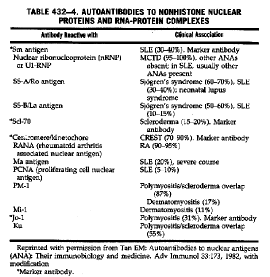
Another, antiribonucleoprotein (anti-M-RNP), has led to the identification of
mixed connective tissue disease (MCTD). Anti-Ul-RNP is also found in SLE, scleroderma,
and polymyositis, as well as in MCTD (Table 432-4). Even more recently, antigens
associated with Sjogren's syndrome-SS-B, an acidic nuclear antigen (cross-reactive
with Ro antigen of cytoplasmic extracts), and SS-A antigen, a similar antigen
(cross-reactive with La cytoplasmic antigen)-have been described. They occur
in about 60 per cent of patients with Sjogren's syndrome and in smaller subsets
of patients with Warker antibody.

SLE (SS-A in 30 to 40 per cent; SS-B in 10 to 15 per cent). SSA (Ro) can cross the placental barrier and is regarded as a marker for neonatal lupus, often with complete heart block. Other significant associations and possibly marker antibodies exist. In scleroderma, the Scl-70 antibody (topoisomerase 1), while present in only 15 to 20 per cent of such patients, is relatively specific. Anti-kinetochore (centromere) antibody occurs in about 30 per cent of such patients but appears in over 90 per cent of those with CREST subset. Antibodies to Jo-1 are present in about 30 per cent of patients with polymyositis but may recognize a subset with interstitial pulmonary fibrosis. The accepted antigens and antibodies and an estimate of their prevalence in disease are listed in Tables 432-3 and 432-4.
In
addition to these, another nuclear antigen, termed RANA, has been found to have
a close association with the Epstein-Barr virus (EBV) and also a high association
with rheumatoid arthritis. Whether this simply represents a marker for rheumatoid
arthritis or implicates the EBV in the pathogenesis of the disease remains to
be seen.
Tests for cell-mediated immunity and for circulating immune complexes (CIC) are of interest, but currently their significance in the rheumatic diseases is not well defined. CIC occur in-infectious diseases, neoplastic states, and glomerulonephritis as well as rheumatic diseases. Methods of measurement are multiple and include the Raji cell assay, C1q binding assay, bovine conglutinin assay, and staphylococcal binding assay. Levels often correlate with disease activity but have no demonstrated value over other rheurnatologic tests and'should not be part of a routine evaluation.
RADIOGRAPHIC TECHNIQUES IN THE DIAGNOSIS OF ARTICULAR DISEASES. Clinical Radiology of Joints. Skeletal radiographs can contribute to the diagnosis of articular diseases because the bone and joint changes reflect the basic pathology. Proper integration depends upon an understanding of the pathophysiology of the various diseases and an appreciation of the typically affected areas. This discussion will highlight general patterns and differential diagnostic aspects of major diseases. The simplest classification is that used in assessing synovial fluid, i.e., inflammatory and noninflammatory articular disease.
General
radiologic features of noninflammatory disease (such as degenerative disease)
include uneven narrowing of
the joint space, sclerosis of juxta-articular bone, and the presence
of bone spurs and cysts. Target areas include the distal interphalangeal and
first carpometacarpal and metatarsophalangeal joints, acrornioclavicular joint,
vertebral column, hip, and knee, with the appearance of genu varum.
Inflammatory joint disease (such as rheumatoid arthritis) is generally characterized by local bony demineralization,'erosions, and uniform narrowing of the joint space. The pattern includes bilateral symmetry with involvement of the metacarpophalangeal joints, u1nar styloid area, and glenohumeral and atlantoaxial joints, as well as metatarsophalangeal joints. Involvement of the knees leads to genu vaIgum.
In addition there are patterns of change that assist in the differentiation of several types of inflammatory articular disease. For example, in tophaceous gouty arthritis the eroded joint may demonstrate an "overhanging ledge," as opposed to the erosion of rheumatoid arthritis. It may show asymmetric intra- and extra-articular erosions, as opposed to the marginal intra-articular erosions of rheumatoid arthritis. Gout does not lead to osteopenia, and joirit space narrowing will be a late phenomenon rather than an early manifestation as it is in rheumatoid arthritis.
In the seronegative spondyloarthropathies (e.g., psoriatic arthritis) the articular involvement is less likely to be symmetrical, the interphalangeal joints of the feet may be more involved than the metatarsophalangeals, osteopenia is uncommon, and periosteal new bone formation may be seen as well as bony ankylosis. Finally, evidence of sacroiliitis with narrowing, erosions, sclerosis, and ultimate obliteration of the sacroiliac joint is common. Descriptions of specific changes, such as calcinosis in scleroderma and polymyositis and aseptic necrosis (osteonecrosis) associated with SLE, are detailed in the chapters devoted to the individual disease entities.
Several additional principles should be stressed in the radiographic examination of joints. First, films are of little value if they are not of good technical quality. Occasionally, for fine points, microradiography (high resolution magnification radiography) can clarify whether or not an erosion or lesion is present. Second, the use of radiography must be selective and usually can be limited both for safety and cost effectiveness. For example, in evaluating rheumatoid arthritis: (1) in the upper extremities, the key films are those of the hands (including the wrists) in posteroanterior and 15 degree oblique views; (2) in the neck, a lateral view in flexion alone will give sufficient data; and (3) in the feet, posteroanterior and lateral views without the oblique will often suffice. To assess degenerative joint disease of the knee, it is vital to include a standing anteroposterior view of both knees in one frame. For examination of the patient with seronegative spondyloarthropathy, limited views of the spine-i.e., a posteroanterior view of the pelvis to visualize the sacroiliac joints, anteroposterior and lateral views of the lumbosacral spine, and a lateral view of the neck in flexion-may suffice.
Joint Scintigraphy-Radioisotopes in the Evaluation of Articular Disease. The use of labeled isotopes in tracer amounts to measure their accumulation over normal and pathologic joints for the assessment of articular disease was introduced in the 1960's. The isotope evaluated first was technetium-99m (1-Tc) pertechnetate, which largely binds to serum protein and is localized in areas of increased tissue vascularity. These scans correlate well with clinical assessment of joints and routine radiography, and on occasion allow the earlier detection of inflammation (increased blood flow). However, they are nonspecific, usually do not add significantly to routine radiography, and add (albeit a small amount) to the total body exposure.
The
introduction of the bone seeking isotope 11-Tc-diphosphonate added a new dimension
to scintigraphy, since. this allowed better evaluation of the axial skeleton.
In many centers this has become the scan of choice. It can on occasion
The routine history, physical examination, selected laboratory studies, and selected routine radiographs make the use of joint scintigraphy a procedure to be chosen by experts for a specific purpose rather than a routine part of the work-up of a patient with arthritis.
Arthrography (Synoviography). The injection of a contrast meditm, often together with air or carbon dioxide, to visualize a joint space has become an accepted diagnostic procedure. Its value lies in the assessment of internal derangements of a joint space (usually a knee or shoulder) rather than in diagnosing inflammatory disease. The usual contraindications are infection or a bleeding diathesis. The radiopaque dye is introduced and the radiograph obtained promptly to determine its dispersion.
Orthopedists use the technique widely (as well as arthroscopy) in the diagnosis of cartilage tears in the knee and rotator cuff injuries of the shoulder. It is of great value medically in assessing masses in the popliteal area. When the differential diagnosis of deep venous thrombosis versus a ruptured popliteal cyst arises, venography is almost always indicated as a prior procedure.
Synovial cysts in rheumatoid disease, e.g., popliteal (Baker's) cysts, may rupture and lead to pain and discomfort about the knee and in the gastrocnernius area. Often the differential diagnosis is between acute thrombophlebitis and ruptured synovial cyst. When thrombophlebitis has been excluded, arthrography can often define the medical problem. On occasion, large synovial cysts can be instrumental in the development of thrombophlebitis caused by the pressure effect. Synovial cysts can occur about other joints but usually are asymptomatic.
Arthroscopy. Arthroscopy is an endoscopic procedure that has widespread orthopedic use. Its major application is in injuries about the knee joint, especially meniscal and cruciate tears. With advancing technology the procedure has also been utilized in other articular spaces.
It is occasionally used by the rheumatologist in undiagnosed monarticular knee disease as a method of obtaining a selective biopsy from a local area of the joint under direct observation. It is not, however, a routine procedure and should only be performed by those with appropriate expertise.
Miscellaneous. Other technologies have been applied to the diagnostic evaluation of articular disease. These include angiography (useful in defining synovial tumors), ultrasonic scanning (increasingly helpful in the assessment of intact popliteal cysts), and, most recently, computed tomography of the joint space. All these procedures have limited utility.
Bartfield H, Epstein WV: Rheumatoid factors and their biological significance. Ann NY Acad Sci 168:1, 1969. A volume devoted to clinical and investigative aspects of the nature of rheumatoid factors and their relevance to disease.
Cohen AS (ed.): Laboratory Diagnostic Procedures in the Rheumatic Diseases. 3rd 6d. Orlando, FL, Grune & Stratton, Inc., 1985. A definitive review of the methodology and interpretation of laboratory tests in rheumatology.
Dixon AS, Rasker JJ: Synoviography. Clin Rheum Dis 2:129, 1976. A fine exposition of symoviography (arthrography) of various joints and the information to be gleaned from these procedures.
Egeland T, Munthe E: Rheumatoid factors. Clin Rheum Dis 9:135-160, 1983. A thorough and up-to-date review of the history of Rl's, their specificity, assay, biologic and pathologic effects, and clinical significance.
Forrester DM, Brown JC (eds.): Radiologic investigation in rheumatology. Clin Rheum Dis 9:289-488, 1983. Eleven papers covering scintigraphic imaging,
computed tomography, sonography, arthrography, and standard radiography in the rheumatic diseases. A British view.
Fritzler MJ: Antinuclear antibodies in the investigation of rheumatic diseases. Bull Rheum Dis 35:1-10, 1985. Excellent review including an algorithm to indicate the diagnostic approach to the use of ANAs in clinical rheumatology.
Goldenberg DL, Cohen AS: Synovial membrane histopathology in the differential diagnosis of rheumatoid arthritis, gout, pseuclogout, systemic lupus erythematosus, infectious arthritis and degenerative joint disease. Medicine 57:239, 1978. An analysis of synovial membrane histopathology in the common inflammatory articular diseases, pointing out patterns that are diagnostically useful even though individual pathognomonic findings are rare.
Hadler NM, Spitznagel JK, Quinet RJ: Lysosomal enzymes in inflammatory synovial effusions. J Immunol 123:572, 1979. A useful study of lysosomal enzymes of synovial fluid that discusses both sides of the question as to whether these enzymes are major causes of articular tissue destruction.
Harris ED Jr, Krane SM: Collagenases. N Engl J Med 291:557, 605, 652, 1974. A scholarly review ofan increasingly important enzyme (coliagenase) that might itself (or through its inhibitor) play a role in cartilage destruction.
Jackson RW, Dandy DJ: Arthroscopy of the Knee. New York, Grune & Stratton, 1976. A simple text that outlines the advantages and hazards of arthroscopy.
Kushner 1, Volanakis JE, Gewurtz H: C-reactive protein and the plasma protein response to tissue injury. Ann NY Acad Sci 389:1-482, 1982. The latest update of the chemistry and significance of C-reactive protein.
Nakamura RM, Peebles CL, Rubin RL, et al.: Autoantibodies to Nuclear Antigens. 2nd ed. Chicago, Arn~rican Society for Clinical Pathology Press, 1985. An authoritative review of antinuclear antibodies and their clinical significance by major contributors.
Ng KC, Brown KA, Perry JD, et al.: Anti RANA antibody: A marker for seronegative and seropositive rheumatoid arthritis. Lancet 1:447, 1980. An interesting test relating to rheumatoid arthritis that may have pathogenetic significance.
Resnick D, Niwayam G: Diagnosis of Bone and joint Disorders with Emphasis on Articular Abnormalities, Vols 1-3. Philadelphia, W. B. Saunders Company, 1981. An extensive review of the radiologic findings in articular diseases.
Ropes MW, Bauer W: Synovial Fluid Change in joint Disease. Cambridge, MA, Harvard University Press, 1953. The definitive work on synovial fluid analyst . s in various articular diseases. The data on synovial fluid white counts, differentials, mucin, clot, etc., of 35 years ago are still valid and represent the basis for most subsequent joint fluid analyses.
Rosenspire KC, Kennedy AC, Russomanno L, et al.: Comparisons of four methods of analysis of 11-Tc pyrophosphate uptake in RA joints. I Rheumatol 7:461, 1980. One of the first critical comparisons of the value and limitations of various methods of joint scintigraphy.