Coronary Artery Disease (or CAD) is the
leading cause of death in the United States, accounting for over 900,000
deaths annually. More than two of every five Americans die of cardiovascular
disease. Additionally, over 3 million Americans suffer occasional chest
pains due to coronary artery blockages. Despite this , the US is still
only 17th in cardiovascular disease mortality worldwide. As common as it
is, CAD has not yet been eradicated by preventative measures. In fact,
it is only within about the last 50 years that the role of cholesterol
and dietary fat in the development of CAD has really been understood. A
number of other "risk factors" have also been identified, including family
history of CAD, hypertension, smoking, diabetes, and lifestyle issues such
as lack of exercise or Type A personality, etc.
The heart is a muscle; like other muscles
in the body, it is composed of millions of specialized cells which contract,
or shorten, under the proper conditions, and develop a force of contraction.
In the heart, the muscle fibers are aligned in a circular manner to form
a conical shaped chamber. As the heart muscle cells contract in unison,
blood is forced out of the chamber and flows to every organ and cell in
the body. This requires a tremendous amount of energy, so the heart must
have its own source of blood to bring nutrients to its cells. A normal
heart in an average sized person will pump 4 to 5 liters of blood per
minute. The average heart will beat almost 4 million times per year.
The "fuel" for this effort comes exclusively from the nutrition carried
to the heart muscle cells by way of the vascular system. This is an image
of a normal heart.
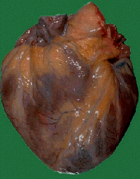
The main chamber for pumping oxygen-rich red blood to the body is called
the left ventricle. After the blood exits the left ventricle, it
enters the main channel of the vascular highway, called the aorta,
a tube of specialized tissue capable of carrying the entire 4 or 5 liters
per minute to the rest of the body under pressure . The very first branches
arising from the aorta are small vessels, called the coronary arteries,
which connect to the surface of the heart. There are two main coronary
arteries, one to the left ventricle (called the left main coronary artery
or LMCA) and another called the right main coronary artery (RMCA) which
supplies mostly the right ventricle but also part of the undersurface of
the left ventricle. These main coronary artery give rise to branches which
thread into the heart muscle, bringing vital nutrients to each muscle cell.
When coronary artery disease occurs, the
channel inside these arteries becomes progressively "plugged" with plaque
material. This process is known as atherosclerosis. In this disease,
the inner channel of the coronary artery becomes obstructed by buildup
of cholesterol and other body fats on the lining of the blood vessel wall.
Over time, the bodies own inflammatory response to these fatty molecules
leads to enlargement and protrusion of the plaque into the flow channel
of the artery. If the plaque is large enough, the entire channel can become
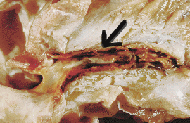
obstructed to blood flow. This is an image of a severely blocked Left Main
Coronary Artery (dissection). You can see the plaque material clogging
the flow channel; it is almost completely blocked on the right of the picture
(arrow).
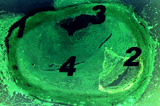
This is a cross-section of a severely blocked artery. (1) is the arterial
wall; (2) is an area of calcified or hardened plaque, intruding into the
cell wall; (3) is the remaining open flow channel; (4) is the fatty material
clogging most of the arterial passage.
When this happens, the heart muscle cells no longer receive vital nourishment
from the blood stream. When the demand for blood supply is increased, such
as during physical exercise and/or emotional stress, the blocked arteries
may not be able to deliver enough blood to meet the needs of the heart
muscle cells. This situation is called ischemia, and is best described
as a shortfall between nutrient demand and supply. As a result, most (but
not all) patients will notice chest pain or pressure with futher exercise.
This pain is called angina pectoris (or just angina). If the obstucted
artery closes suddenly, then permanent damage to the muscle cells in that
region of the heart can occur. This process is known as a heart attack,
or myocardial
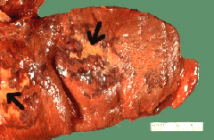
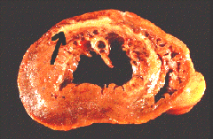
infarction (MI). These two images show the damage from a severe
MI; the pale tannish tissue (arrows) has died and formed scar tissue. In
the second image, a cross-section, the area of infarction extents from
the ventrical wall into the septum, or wall between the left and right
ventricals.
Some patients will need intervention to prevent
further attacks. Surgery is not always necessary, but for many patients
surgical revascularization still provides the best long term result.
Diagnosis: Angiography
Coronary Angiography is the diagnostic
x-ray procedure designed to visualize the arteries of the heart. These
tiny blood vessels are only 1 to 3 millimeters in diameter. Thus it takes
special x-ray equipment and techniques to obtain images of sufficient quality
for diagnosis and surgical decision making. Coronary angiography has become
the cornerstone for both diagnosis and treatment of coronary artery disease
(CAD) worldwide.
In this test, the patient is brought to the
x-ray (or radiology) procedure room where specialized fluoroscopic equipment
is available. The patient must lay motionless on the x-ray table, with
their hands to the side or overhead. It is normal for a patient to be apprehensive
and nervous during this procedure. The team in the catheterization laboratory
(or cath lab) will commonly administer medications designed to relax the
patient and relieve both pain and anxiety during the procedure. A sterile
area is prepared in the groin by cleansing the skin with topical antiseptic
agents and covering the region with sterile drapes. The skin is "numbed"
with local anesthesia (similar to the Novocaine injection used by a dentist).
The cardiologist places a needle into the main artery in the upper leg,
called the femoral artery. Through this needle, a flexible wire is passed
and threaded backward into the arterial tree until the wire reaches the
main aorta (as confirmed by x-ray fluoroscopy). Over the wire, a long plastic
tube (also known as angiogram catheter) is threaded into the aorta. The
initial wire serves as a guiding system to ensure that the catheter tracks
into the aorta properly.
The angiogram catheter is maneuvered into
position just above the outlet valve of the left ventricle, (also known
as the aortic valve). With careful maneuvering, the tip of the catheter
can be positioned at the mouth of the main coronary arteries. Once positioned,
specialized contrast agent, or angiogram dye is selectively injected into
the mouth of the coronary artery while continuous x-ray images are exposed
onto movie film. During the actual injection, the patient notices a flushing
sensation starting in the chest and migrating to the head and sometimes
the entire body. This sensation is a side effect of the dye matierial,
and occurs to a varying degree in all patients. The flushing sensation
is bothersome, but is short-lived. It is not an allergic reaction.
" Thallum images:
A. On planar images, there is borderline increased lung uptake.
There are severe perfusion defects involving the anterior, septal, apical
and inferior walls. The lateral wall is relatively preserved. The cavity
is enlarged. On delayed images, there is some degree of redistribution
in the septum and inferior wall.
B. On tomographic images, there is a severe, primarily fixed anteroseptal
and apical perfusion defect that partly distributes at 3 hours. The lateral
wall is also abnormal at peak exercise, and significantly redistributes
on delayed images.
Left ventricular function was severely impaired with evidence of
an anterior septal and apical aneurysm. "
As the dye travels down the branches of the
coronary artery itself, continouous images are exposed onto movie film.
The composite roll of images is known as a cineangiogram. In most modern
cath labs, the cardiologist can also see the preliminary results immediately
on the overhead video screen. Digitized images are also saved on computer
and replayed onto the video screen as needed. In nearly all cases, multiple
views from different angles are necessary in order to ascertain the precise
location and severity of the coronary artery blockages. In some views,
a blockage may not initially appear to be that severe. However, from another
angle of view, the artery may appear nearly occluded. Thus it is important
for the cardiologist to rotate the fluoroscopy equipment and obtain pictures
from multiple views.
After completion of the coronary angiogram
procedure, the cardiologist reviews the final images on a specialized projector,
making measurements of each blockage seen. Blockages in the channel of
an artery are rated by percentage (i.e. the percent of the flow channel
that is obstructed). For blockages that involve less than 50% of the vessel
diameter, intervention is usually not necessary. In general terms, when
plaque buildup is blocking more than 50% of the vessel diameter, the blockage
is considered clinicially significant. However, most blockages are not
considered serious until they reach at least 75% narrowing of the diameter
of the artery. At this point, there will always be significant impairment
of blood flow. Such lesions are clinically important and thus candidates
for treatment.
This is an example of a 99% blockage in the most important coronary
artery, the left anterior descending (LAD). The normal parts of the coronary
vessels are clearly seen as smooth unbroken white channels against the
black background of the undyed tissues.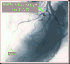
Here is a view of the right coronary artery
(RCA) demonstrating two high grade (> 90%) blockages.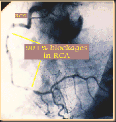
In my case, I was diagnosed as having "severe three-vessel" CAD; none
of the three main arteries were operable, and th e damage had already been
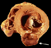
done. In addition, as a result of the MI, I had an aneurism at the base
of my heart, somewhat like the one shown in these images. In an aneurism,
a part of the heart wall bulges out and becomes very thin and non-contractile.

Because most of the damage was old, the stress placed on the remaining
functional heart muscle of my left ventrical had caused it to enlarge and
become "flabby", resulting in Congestive Heart Failure, a condition in
which the heart is not able to pump the blood with sufficient energy to
carry fluids out of the tissues of the body, especially the lungs, lower
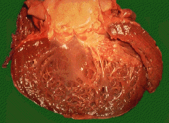
extremeties, and around the heart. This is an image of a severely enlarged
heart, showing the left ventrical; compare this to the normal heart above.
Depending on lesion size, shape, length,
location and other factors, some blockages can be treated without bypass
surgery, by techniques like baloon angioplasty. However, there are many
patients in whom the blockages cannot be easily or safely modified by such
techniques. Coronary Artery Bypass Grafting (CABG) is the treatment of
choice for such patients. For example, in patients with multiple high grade
blockages, it is more effective to restore circulation throughout the heart
with bypass surgery. If the cardiologist feels that surgery is warranted,
the films and clinical situation are reviewed by a consulting surgeon.
The surgeon utilizes the cineangiogram images to plan both the location
and number of potential bypass grafts needed in order to optimize that
patients surgical result.
"Films were sent for review. Dr. Sansonetti felt that the
patient was a surgical candidate and agreed to accept the patient for further
evaluation. The patient is transferred and coronary bypass graft surgery
is anticipated for the next several days.
Transfer Diagnoses:
Coronary artery disease with probable prior, clinically unrecognized
anterior septal myocardial infarction with extensive remodelling, aneurysm
formation, moderate-to-severely impaired left ventricular function, but
with normal diastolic pressure and absence of congestion. Recent unstable
angina with severe three vessel coronary artery disease."
Any surgeon intending to reconstruct the circulation
to the heart will need a road map of the blood vessels and the location
of the blockages. The test most frequently used for this information is
called a coronary angiogram. In this test, the small blood vessels
feeding the heart are imaged with special x-ray techniques. In summary,
the coronary arteries are injected with a special dye solution, usually
a slightly radioactive thalium solution, which shows up on x-ray film.
As the dye travels down the branches of the coronary artery itself, moving
pictures (or cineangiograms) are taken with the x-ray camera. The patient
can sometimes watch this on a video screen. The series of angiographic
movies are recorded and reviewed by the cardiologist and decisions then
are made about the relative benefits of different treatments.
Surgery
Surgical reconstruction of the blocked arteries
began in the late 1960's and has proven to be extremely successful. The
operation called Coronary Artery Bypass Grafting (CABG or "cabbage") is
the foundation of surgical management of heart disease, restoring the blood
supply to the heart muscle by creating a new route (aka bypass) for the
blood to flow around the blockages. CABG is the most common operation performed
on the human heart, and in fact is currently the most common procedure
of any kind in the USA. Last year, over 200,000 CABGs were performed nationwide.
Now there are many other treatment options,
but surgery remains the mainstay for a large group of patients for which
other options are not effective. Coronary Artery Bypass Grafting doesn't
remove the obstructing blockages in the arteries. The procedure "reroutes"
the flow of blood by using a "detour" pathway. Typically, the blockages
occur in the first centimeter or two of the major branches feeding the
heart. The smaller branches are usually not involved until very late life.
Thus, it is possible to hook a new source of blood into the artery just
beyond the last major blockage. Blood flows into the artery through a different
path (such as a vein bypass graft) and reaches the heart muscle tissue
where it is needed. Once the blood flow is restored, the symptoms of exertional
chest discomfort are relieved .
"This 47 year old patient entered St. Vincent Hospital with
chest pain and syncope. He has a reduced EF. The circumflex is the only
artery open and it has 95% lesion. The anteroapical wall is profoundly
dilated and akinetic."
The most common material used to build this
new pathway is a vein from the lower extremity called the Greater Saphenous
Vein (or GSV) running from just inside the ankle bone up to the groin.
This vein is the right size, shape, and length for use as a bypass conduit.
The other major vessel used as a bypass graft is the Left Internal Mammary
Artery (LIMA),which lies alongside the undersurface of the breastbone (aka
sternum). By detaching the lower end of the LIMA, the vessel can be transplanted
to the surface of the heart.
"With the patient supine on the operating table, general
endotracheal anesthesia was induced and the patient was prepared and draped
sterilely from the chin to the ankles. The greater saphenous vein was harvested
from the left thigh. It was good caliber and quality. The side branches
were ligated with 4-0 silk and when the incision was dry it was closed
in the usual fashion."
Regardless of which conduit is used for the
bypass, the main advantage offered by this surgical approach is the restoration
of blood supply to the heart. The vein or mammary artery provide a new
and unobstructed route for blood flow. When the surgeon connects the bypass
to the native coronary artery beyound the obstruction, blood has a new
path in which to flow into the blood vessel beyound the blockage. Then
when the heart demands more blood flow, the situation can be met by the
new source of blood flowing through the bypass graft. It is important for
the surgeon to provide a bypass for each major branch that is obstructed.
In that way, the whole heart will have more blood supply, and the anginal
(chest) pains will be relieved.
A typical CABG operation begins with a vertical
opening into the front of the chest. The breast bone is divided with a
specialized (reciprocating) saw, similar to a "jig" saw used in woodworking
designed just for this incision. The split sternum is spread open with
a device that functions like a winch. The soft tissues in front of the
heart are parted, and the membrane surrounding the heart (the pericardium)
is incised. Next, the surgeon removes the Internal Mammary Artery from
the chest wall to provide a donor artery for grafting.
Simultaneously, an assistant surgeon proceeds
with harvesting additional conduit, usually the Greater Saphenous Vein
(GSV) from the inside of the calf or thigh. This vein is long, straight,
and the proper caliber for use as a donor graft to the heart and removal
of the GSV does not impair the return of blue blood from the extremity.
"Simultaneously, a median sternotomy was perfomed, the sternum
was split with an oscillating saw, the sternal retractor was placed. The
pericardium was then opened and the patient was sytemically heparinized.
The aorta was cannulated in the usual fashion and a single venous cannula,
two-stage, was placed through the right atrial appendage. A cardioplegia
needle was placed proximal to the aortic cannula and was secured."
After all the donor vessels are harvested, the
patients blood is thinned with a large dose of the potent anticoagulant
heparin. This renders the blood incapable of clotting when exposed to the
plastic tubing and surfaces of the heart-lung machine. The patients circulation
is connected to the heart-lung bypass circuit, and the body placed onto
the support machinery. Then the body temperature is lowered (by refrigerating
the blood as it circulates through the machine). In addition, blood flow
to the heart is separated from that going to the rest of the body by a
vascular clamp applied to the aorta just below the insertion of the arterial
return cannula. The coronary arteries are then perfused with a cold potassium
solution. The heart is immediately rendered motionless, cold, and relaxed.
The body is preserved by the nutrient flow provided by the heart-lung circuit,
while the heart is preserved by the low temperature and other conditions
managed by the surgeon.
"Cardiopulmonary bypass was initiated and the patient was
kept warme while a retrograde cardiplegia catheter was placed through the
right atrium into the coronary sinus. The paient was then cooled to 27degrees
C. When the heart arrested from the cold, the aortic crossclamp was placed
and 400cc of cold blood cardioplegia were infused into the aortic root
and 400cc's were infused retrograde into the coronary sinus. There was
good electrical arrest of the heart and the heart was well decompressed.
Iced saline was placed in the pericardial sac."
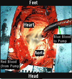
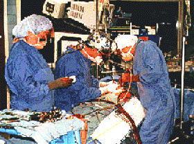
These are images of the procedure, with the heart-lung bypass machine attached
to the heart.
Next, each target vessel is identified as it runs across the surface
of the heart. For each intended bypass, the surgeon makes a tiny opening
into the front wall of the target coronary artery with a very fine knife.
The opening is expanded with specialized scissors. A donor vessel, either
vein or IMA is stitched to this opening with delicate fine suture material.
Currently, the best suture material is made from polypropylene and is finer
in diameter than a human hair. After all the grafts are sutured to the
heart arteries, the vascular clamp is released, allowing blood to flow
into the native heart arteries again.
"The heart was inspected to see if there were graftable
arteries that did not show up on the cadiac catheterization. A large area
of the apex was a thin scar that inverted when the heart was well decompressed
and the entire ridge along the LAD was dense and infarcted. The right coronary
was inspected. It appeared non dominant. The OM1 was an intramyocardial
branch. It was thin-walled and not easy to work with."
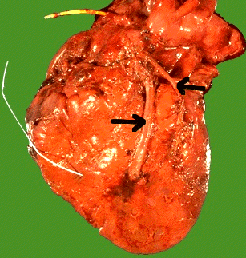 Internal
Mammary Artery grafts are already attached at their origin from the main
artery to the arm (the subclavian artery). Blood flowing through the IMA
directly comes from the subclavian artery. However, GSV grafts are detached
at both ends. After connecting one end of the vein to the coronary artery,
the other end must then be connected to a source of red blood. This is
done by partially occluding a segment of the ascending aorta with a specialized,
curved vascular clamp. Holes are created in the front wall of the aorta,
and the veins are anchored to these openings with fine suture. After releasing
this partially occluding clamp, the veins fill with red blood from the
aorta and deliver this blood to the coronary arteries downstream. This
image shows a heart with two grafts (arrows); the wire is for a pacemaker
device, sometimes needed if arrhythmias are present.
"An end-to-side anastomosis was performed. The flow was
80 cc's/min. Cardioplegia was infused through the graft and through the
root. An attempt was made to re-infuse the cardioplegia retrograde, but
the coronary sinus catheter, despite being deflated when the heart was
being moved, had poked out through the coronary sinus and the retrograde
catheter was then pulled back a little bit and the rent in the coronary
sinus oversewn with 4-0 prolene. The retrograde catheter was removed and
furher cardioplegia was given through the graft and through the root. There
was some electrical actiity during the rest of the graft. The true inferior
wall of the heart was served by what appeared to be the OM-2 branch. An
end-to-side anastomosis was perfomed and the flow was 180 cc's/min."
After the heart has had a chance to recover
from the temporary arrest period, the heart-lung circuit can be gradually
withdrawn. When the heart is beating strongly again, then the heart-lung
machine is stopped, and the equipment removed. The anticoagulant (heparin)
is chemically reversed (with a drug called protamine). The surgeon then
inspects and controls any remaining bleeding. Finally, the wounds are closed
and the patient sent to intensive care for recovery.
"The aortic crossclamp was removed. The patient was slowly
rewarmed to 37 degrees C. The heart started beating ventricular tachycardia
requiring two cadioversions of 30 joules to achieve normal sinus rhythm.
The cardioplegia needle was removed and a Lambert Kay clamp was placed
on the aortic root. The cardioplegia site was used for one proximal anastomosis
and a 4.5mm punch was used to make one additional aortotomy site for the
the saphenous vein grafts. These were performed in an end-to-side fashion
using 5-0 prolene running suture. Marker rings were applied and attached
loosely to the proximal anastomotic area using the same prolene suture.
Hemosatis was adequate at all anastomotic sites. Two right ventricular
pacing wires and two atrical appendage wires were placed and secured. Because
of the patient's preoperative cardiac index of 1.5, and the known dilated
cardiomyopathy, 5 mcg. of Dobutamine was initiated. There was good contractility
response to this, and the patient came off bypass well with good BP, normal
sins rhythm of about 100, and as the case progressed, the Dobutamine was
weaned to 2.5 mcg. When hemodynamics were stable, the venous line was removed
through purse string suture and Protamine was given to reverse the Heparin.
The arota was decannulated and a horizontal mattress suture of 4-0 prolene
was used in addition. Two straight #36 chest tubes were placed in the pericardial
space. Mediastinal fat and some of the pericardium was closed anteriorly
with a 3-0 prolene. The sternotomy was closed with heavy wire and two layers
of Dexon in the usual fashion. Sterile dressings were applied and the patient
was transported to the Intesive Recovery Room in stable condition."
Many patients, without any major post-operative
problems, can leave the hospital in about 4 to 6 days from surgery. It
will take about another several weeks for most patients to feel stronger
and regain normal body habits, such as appetite, sleep patterns, bowel
patterns, etc. For patients who work in non-physical jobs, many can return
to employment within 4 to 6 weeks of surgery. Usually, antianginal medications
are no longer needed. However, high blood pressure, diabetic medications
, cholesterol-lowering drugs and other medicines (such as ACE inhibitors
and Beta Blockers) will often be prescribed to treat the underlying causes
of the disease. A program of exercise and diet regulation is also usually
prescribed, as well as stress-reducing programs and, often, counselling.
Other Information
Glossary:
-
Ace Inhibitor
A heart medication of complex chemistry which reduces the heart's workload
-
Aneursim
A bulging of the heart or artery wall outward, with thinning of the
tissues and often, scarring. Can produce a thrombus.
-
Angina
The chest pain associated with inadequate blood supply to the heart
-
Angiogram
A diagnostic procedure to determine extent of arterial blockage
-
Angioplasty
A procedure in which a catheter is inserted through the blood vessels
to the constricted arteriy and then a "baloon" is inflated inside the artery
to open it.
-
Aorta
The main blood vessel leading from the heart and connecting to the
"vascular tree"
-
Atherosclerosis
" Hardening of the arteries"
-
Beta Blocker
A drug which limits how hard the heart can work
-
Cardiomyopathy
Death and/or damage to heart muscle cells
-
Cardiovascular Accident
A blood clot or thrombus which breaks loose and causes damage in some
other part of the body (often the brain, resulting in a stroke)
-
Cholesterol
Essential fatty proteins used by the body in making hormones, etc.
Occurs in food only in products derived from animals. There are two kinds;
LDL (Low Density Lipoproteins) - the "bad" kind, and HDL (High Density
Lipoproteins) - the "good" kind, which function as "sweepers" to remove
LDL cholesterol from the body via the liver and gall bladder
-
Cholesterol reducing drugs
Drugs which act on the liver to reduce the natural production of cholesterol
by the body
-
Congestive Heart Failure
Progressive failure of the heart's pumping ability, resulting in accumulation
of excess fluids, especially in the lungs; can result from severe heart
attack
-
Coronary Artery Bypass Graft (CABG)
Surgical procedure in which blocked arteries are "bypassed" by grafting
veins to resupply the heart muscle with blood
-
Diuretic
Drug to assist in the elimination of excess fluid buildup
-
Echocardiogram (see Sonogram)
-
Edema
Swelling due to abnormal accumulation of fluids in tissues
-
EKG
Electrocardiogram; a diagnostic tool that measures various activities
of the heart
-
Fibrillation
Rapid but shallow heart beat
-
Heart Attack
Caused by inadequate blood supply to the heart muscle resulting in
death of heart muscle tissue
-
Hypercholesteremia
Abnormally high serum cholesterol coupled with low percent of HDL (good)
cholesterol
-
Hypertension
High blood pressure which can cause thickening (hypertrophy) of the
heart walls
-
Infarct, Infarction
Death of cells
-
Lipids
Fatty proteins either from dietary sources or produced naturally by
the body
-
Myocardial Infarction
A heart attack
-
Pacemaker
An electrical device to replace the natural pacing mechanism when it
no longer functions properly
-
Cardiac Rehabilitation
The process of recovery after a heart attack, surgery or angioplasty,
usually combining an exercise program, diet and psychological counselling
-
Saphenous Vein
A long vein in the legs harvested during CABG surgery for grafting
-
Sonogram
An imaging technique using ultrasound, similar to that used for pregnant
women
-
Stress Test
A diagnostic test involving walking on a treadmill while thallium is
injected into the blood stream, followed by X-rays and/or sonograms.
-
Stroke
A "brain attack," causing death of brain cells due to blockage of blood
supply
-
Thrombus
A blood clot, which often forms following damage to heart muscle
-
Transfat
A type of fat resulting from the hydrogenation of oils, especially
vegetable oils; used in many foods, manufacturers are not required to list
transfat content. Greatly increases the actual total saturated fats.
-
Vasodilator
A drug which relaxes smooth muscle tissue and opens blood vessels
-
Ventrical
The left chamber of the heart
Signs of a Heart Attack:
-
Pain, "heaviness" or crushing sensation in center of chest, sometimes radiating
to the back, the neck and jaw and the left arm. Not related to exercise
or other exertion nor relieved by resting.
-
Heartburn-like sensation not relieved by antacids or other remedies
-
Faintness, dizziness, cold sweat
-
Shortness of breath on normal activity
These symptoms may or not signal a heart attack, but if they continue or
are recurring, you should see your doctor as soon as possible. A large
percentage of people do not survive their first heart attack. In my case,
I had had two major heart attacks and not known it, so-called "silent"
heart attacks. It is important to know your family's medical history and
if you are genetically susceptible to heart disease, these symptoms may
be a serious danger sign and should be taken seriously. An unneeded trip
to the emergency room is better than a trip to the morgue!


 Internal
Mammary Artery grafts are already attached at their origin from the main
artery to the arm (the subclavian artery). Blood flowing through the IMA
directly comes from the subclavian artery. However, GSV grafts are detached
at both ends. After connecting one end of the vein to the coronary artery,
the other end must then be connected to a source of red blood. This is
done by partially occluding a segment of the ascending aorta with a specialized,
curved vascular clamp. Holes are created in the front wall of the aorta,
and the veins are anchored to these openings with fine suture. After releasing
this partially occluding clamp, the veins fill with red blood from the
aorta and deliver this blood to the coronary arteries downstream. This
image shows a heart with two grafts (arrows); the wire is for a pacemaker
device, sometimes needed if arrhythmias are present.