Dr. Dave's Museum - "Whazzat Thang?" - Ole Thangs
Solutions to Past Thangs
Thang #1 |
Thang #2 |
Thang #3 |
Thang #4 |
Thang #5 |
Thang #6
 Unknown Thang #5 (June-August 98)

 Unknown Thang #5 (June-August 98)
Guess(es) Received
It's Teddy & Yatsu here. Our guess to "Whazzat Thang?" is that it's a colony counter for Petri dishes.Am I right?
Teddy Lim, Toronto ON
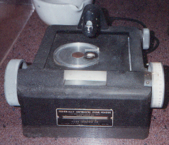
Date: Fri, 28 Aug 1998 19:15:07 -0400================== A N S W E R ================== You're close, but a piece from the microbiology laboratory again proves to be a stumper. This is an antibiotic zone reader, made by Fisher-Lilly. (Those of you with high-resolution monitors can probably read the label on the front!)
Why It Is Needed
When treating infectious diseases caused by bacteria, it is important to use antibiotics that the pathogen is sensitive to, i.e. that will kill it. To determine which drugs those are, microbiologists use a method called Kirby Bauer Antibiotic Sensitivity Testing. Briefly, a sample of liquid culture containing the bacteria in question is uniformly plated out on solid agar medium in a Petri dish. Small cardboard discs impregnated with known amounts of different antibiotics (one per disc) are placed on the surface of the medium, and the plate is incubated. The bacteria multiply, and the antibiotics diffuse out of the discs into the agar. Eventually the bacteria form a confluent mass ("lawn") covering the surface of the medium -- except in areas where there is a lethal concentration of an antibiotic to which they are sensitive, the zone of inhibition. To quantitate that sensitivity, the size of the zone must be determined. A wider zone around a disc means a proportionately greater bacterial sensitivity to the corresponding drug (the bacteria die further from the disc, where the drug concentration is lower). For some antibiotics, bacteria won't be deemed "sensitive" unless the zone is a minimum size. That's where the zone reader comes in.
How It Works
The zone reader basically uses an optical method: After incubation, the plate is placed on the reader surface. A light beam comes up through the hole in the centre of the base, aimed at a mirror at the end of the arm overhanging the reader surface. The beam will pass through the plate only in an area where no bacteria are growing, such as the zone of inhibition. The mirror reflects the beam to the front of the reader, where a calibrated scale (the clear window) allows the size of the zone to be accurately measured.
Devices like the one shown above are actually still in use. They look a little snazzier, but the principle behind their operation is the same. I'll let the manufacturer explain in detail how it works (see the catalogue entry for the Fisher Lilly Antibiotic Zone Reader II).
Simpler measuring devices like rulers and calipers (fancy rulers) are also used. Newer developments in antibiotic sensitivity testing include automated systems that can rapidly read multiple zones, and, in a complete departure from the Kirby Bauer method, a technique that exploits the increased permeability of antibiotic-damaged bacteria membranes to the fluorescent dyes of the BacLight Viability Kit from Molecular Probes Inc., detected by flow cytometry.
With the increasing emergence lately of "super bacteria" having multiple antibiotic resistance, antibiotic sensitivity testing is ever more important.
"Whose Planet Is It Anyway?"
Information on a CBC Radio series about the superbugs with good links (scroll to the bottom)
Unknown Thang #6 (September - December 98)

Guesses Received:Over four months and no guesses! Not enough old hematology technologists surfing the 'Net, I guess.
================== A N S W E R ==================
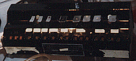
This is a white blood cell differential counter, or more correctly "count keeper", in that it does not count the cells itself but keeps track of the number of cells counted by the hematology technologist.The most basic definition of the field of pathology is "the study of human tissue and body fluids for the purpose of detecting disease." Doctors can learn a great deal by studying a patient's blood, a tissue in liquid phase that is readily sampled with the poke of a needle. Blood is made up of three types of cells:
There are many different types of white blood cells, each with a unique microscopic morphology. When the total number of white blood cells ("white blood cell count" or simply "white count") is increased, doctors want to know the absolute and relative quantities of the different types of white cells, since the resulting profile will give them a clue about the type of disease the patient has.
- red blood cells ("red cells" or erythrocytes), which contain hemoglobin and carry oxygen through the body
- platelets, which are involved in the blood clotting process
- white blood cells ("white cells" or leukocytes), which fight infections
Here's How It Works
The hematology technologist performs a differential white blood cell count (or simply "differential") by examining the blood under a microscope and classifying and counting each white cell encountered. Each type of white cell has its own key (the silver things), which when pressed increments the counter for that type (the numbers below the keys and the white labels bearing the names of the cell types) and the overall count (the red numbers at the left end). When the overall count reaches 100, a bell goes off. The technologist stops counting and transcribes the numbers to a report.
Like typists of the day, technologists back then must have developed strong fingers (in one hand)! Nowadays, the process of doing differentials is automated, so technologists have been spared this tedious work (and, in some cases, have lost their jobs). However, occasionally a manual differential is needed (low sample volume, low cellularity, Y2K ;-) ), so devices similar to this are still in use, with new interfaces like LED displays and LCD displays with hookups to laboratory information systems.