Site Features
 Wingo
Bio Wingo
Bio
 Alaska Alaska
 Bavaria Bavaria
 Russia Russia
 South
America South
America
 Special Forces Reunion Special Forces Reunion
 Photography Photography

Memorial to Chad
|
Case Report
A 23 year old Black Male soldier presented to the US ARMY
Dental Clinic Schweinfurt, Germany with a history of swelling and pain
in the area of tooth #8 (Maxillary anterior tooth). The patient had been
placed on Pen Vee K 500mg qid 4 days prior. Clinical examination revealed
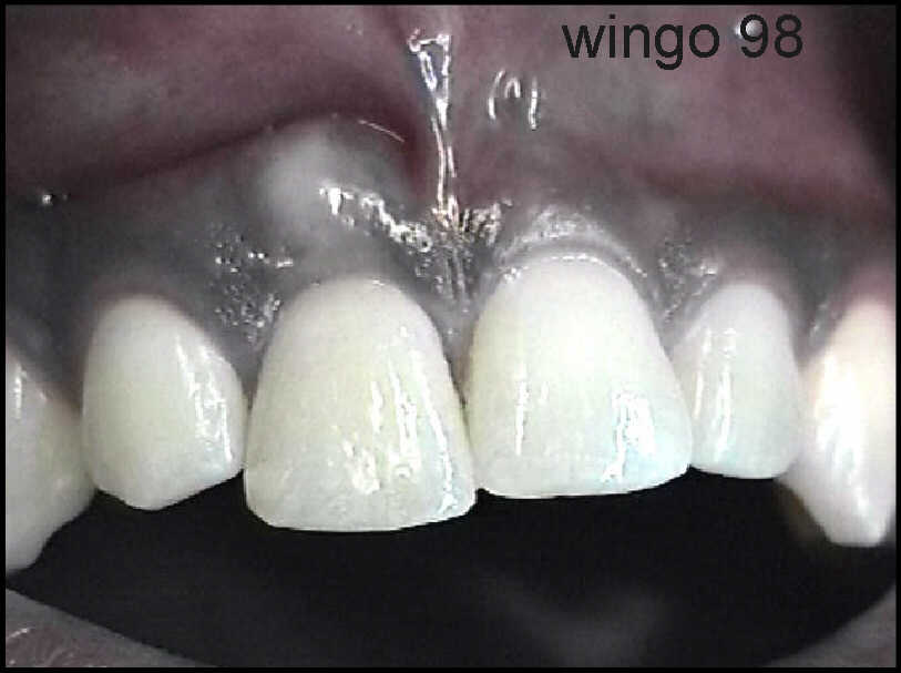
a raised white area over the root of tooth #8 and patient admits to having
been struck in the mouth, 9 months prior, while playing basketball. Additionally
the patient gave a history of previous endodontic therapy to tooth #8.
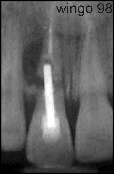
 A PA-Xray was taken demonstrating
an obvious root fracture, and associated periapical radiolucency to include
verification of Endodontic Treatment consistent with history given by the
patient. A metal post had also been placed in the tooth for added support.
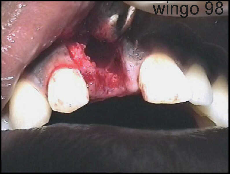
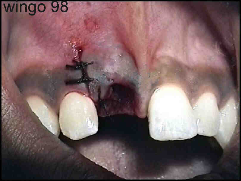
 Treatment of choice was to
remove the Clinical tooth and root. Thirty six mg. of Xlyocaine and
.018mg of epinephrine was administered via local infiltration. A vertical
incision was made and a full thickness flap was reflected exposing bone
and residual root. Bone was removed and the root elevated.
 Associated pathology
was removed, bone smoothed, and the flap was repositioned and sutured with
3 (4-O) silk sutures.
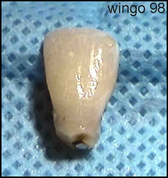
 The clinical crown was
thoroughly debribed of granulation tissue and cleaned.
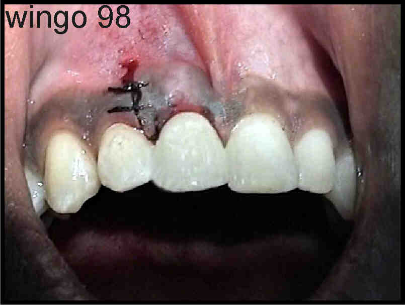
 The teeth on either side
of the removed tooth, to include the clinical crown which had been removed,
was etched with phosphoric acid, washed and air dried. A sealer was applied
to the etched surfaces and cured with Ultra Violet light. The tooth was
repositioned in the socket and Filled-Light-Cured resin was applied to
the interproximal spaces and cured.
The finished result
was an immediate, temporary bridge, using the patient's natural tooth and
little time was necessarily spent reducing adjacent teeth and fabricating
a polycarb bridge. Additionally, unnecessary tooth structure was lost and
conservative treatment was accomplished paving the way for a Maryland Bridge.
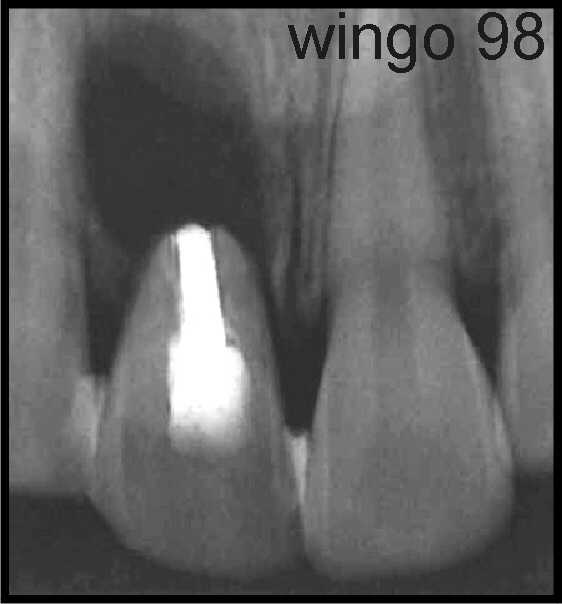
 The post operative radiograph
demonstrates the natural tooth in place. It is expected that the patient
will receive final treatment in 4 to 6 months, hence, allowing sufficient
time for the wound to heal.
Dr. Harold H. Wingo
US ARMY Clinic
Schweinfurt, Germany |

