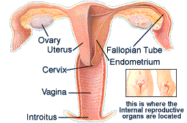

Internal
Anatomy
Vagina (Vuh-JEYE-nuh)
The vagina, also known as
the birth canal, leads to the internal reproductive system. It is
a narrow tunnel that measures between three and five inches in
length. The vaginal opening is called the introitus. The vagina
is surrounded by muscle and supporting tissue that can expand
enough to allow passage of a baby. The natural vaginal secretions
provide lubrication and keep a healthy balance of bacteria in the
vagina to resist infection.
A certain degree of vaginal discharge is normal, and may change
in consistency depending on the hormones present at different
stages of the menstrual cycle. For example, during mid-cycle,
called ovulation, when estrogen levels are high, the cervix
produces a large amount of watery secretions that you may
perceive as vaginal discharge. Normally, it looks clear and
stretchy, and feels slippery -- like mucus. Nature's aim during
ovulation is to enhance reproduction: the mucus is abundant and
slippery, giving sperm the best chance possible of surviving in
the cervix. In contrast, cervical mucus becomes thicker or
disappears entirely at other times of the menstrual cycle.
After menopause, and even the few years leading up to menopause
and thereafter, vaginal discharge may decrease. This reduction in
lubrication, which is caused by lower levels of estrogen, may
cause dryness, irritation or even infection.
Cervix
(SER-viks)
At the top of the vagina is the cervix, or the connection between
the vagina and the uterus. The cervix itself is only about an
inch in diameter, small and pink. The opening to the cervix,
called the os , is a very small hole in the middle of the cervix.
After pregnancy, it appears as a 1/4-inch slit. The os opening is
big enough to allow the flow of fluids, such as menstrual blood,
from the uterus. During the labor of pregnancy, the os opens to
9–10 centimeters to allow delivery of the baby. In
non-pregnant women, the os is only open a few millimeters.
Cervical mucus acts as a barrier to bacteria by preventing
bacteria from entering the uterus and the fallopian tubes.
Many women notice changes in the consistency of the cervical
mucus during their cycle. It is often thin and mucus-like during
ovulation when estrogen levels are high, and thicker and more
sticky -- or seemingly nonexistent -- at any other time of the
menstrual cycle.
Uterus
(YEW-ter-us)
The uterus is a rose-hued, pear-shaped, muscular organ that can
expand and stretch enough to accommodate the development of a
fetus. The inner lining of the uterus is called the endometrium,
which is a lining made up of blood vessels, specialized glands
and supporting tissue. This is the part of the uterus that is
shed during a period.
There are three openings to the uterus. The cervix, which is the
lower part of the uterus that opens to the vagina, and each
fallopian tube which enters the uterus towards the top, one on
either side.
The main function of the uterus is to create a nurturing
environment for the growing fetus. During pregnancy, this small
mass of muscle starts out about the size of a pear, and grows to
become the largest muscle in the body, larger even than thigh
muscles.
During menstruation, the uterus may contract in response to a
series of hormonal changes: the shedding endometrium releases
prostaglandins, which trigger contractions. Furthermore, the
decline in progesterone that occurs just prior to menses may also
contribute to uterine contractions. During labor, uterine
contractions thin the lower segment of the uterus and cervix, a
process called effacement, and expand the cervical os to prepare
for delivery of the baby (dilation). Uterine contractions also
assist in the actual delivery of the baby.
At menopause, as estrogen levels fall off, the endometrial lining
no longer sheds and menstruation comes to an end.
Fallopian
tubes
Besides the cervix, there are two other openings in the uterus
leading to two fallopian tubes. These soft, limp tubes extend
about five inches from the uterus to the ovaries. There are four
components that make up the fallopian tubes: the first is the
intramural component, which is the segment that goes through the
uterine muscle. The second component is the isthmus , or the
first part of the tube after exiting the uterus. The next
component is the ampulla . This is where fertilization occurs.
Finally, the fallopian tubes end at the fimbria , which are
fringed and trumpet-shaped with minuscule feather-like tissue at
the end which grasp eggs (ova) that are released from the
ovaries.
From a reproductive standpoint, the fallopian tubes are designed
to perform four related functions: connect the ovary and the
uterus; transport sperm in the right direction (from the
fallopian tube toward the uterus); provide a meeting place where
conception happens; and, help propel the fertilized egg by
producing gentle, continuous contractions that move the egg
toward the uterus.
Ovaries
(OH-ver-eez)
The fallopian tubes lead to the ovaries which are oval-shaped
organs that secrete hormones and house eggs, or ova. Measuring
about an inch and a half wide and an inch long, the two ovaries
sit on either side of the uterus, attached to the uterus by a
ligament.
The ovaries can be smooth, or during ovulation, they become
marked by clusters of rounded bumps, or follicles , which house
and nurture eggs. The number of eggs that are contained in the
ovaries depends on the age of the woman. The highest number is
actually found before a girl is born. While still in the mother's
womb, a 20-week-old female fetus has approximately 7 million
eggs. At birth, the number has decreased to 2 million. By the
time a girl enters puberty, she has between 300,000 and 500,000
eggs. This decline in number is the process called atresia , a
natural and continuous process that is uninterruptable. Only
between four and five hundred will ripen into mature eggs during
a lifetime.
During the first half of the menstrual cycle, the follicles are
growing and secreting estrogen and the egg is undergoing the
maturation process. The egg continues to grow until it is
released from the follicle and picked up by the fimbria and
transported to the fallopian tube. Meanwhile, the empty follicle
cells coalesce into a yellow mass called the corpus luteum ,
which secretes estrogen and progesterone. Progesterone is
produced to support the gestation (or nurturing) of an egg in the
event that it is fertilized and implanted in the uterus. If
pregnancy does not occur, the estrogen and progesterone secretion
from the corpus luteum will cease 11-16 days after ovulation.
Without the support of the hormones, the endometrium will shed.
Over time, the corpus luteum becomes incorporated back into the
ovarian tissue.
As menopause approaches, ovarian estrogen begins to decline.
Estrogen levels become very low once there are no remaining
follicles in the ovaries. Without ovarian production of estrogen
and progesterone, the endometrium is not stimulated to grow and
shed. This eventually leads to the end of menstruation.
[Homepage] [External Anatomy] [Menstrual Problems] [Introduction] [Breast Self-Exam]
The contents of this Web site are for informational purposes only and are not intended to be used for medical advice. You should consult your physician or health care provider on a regular basis. You should consult your physician immediately with any problem about which you are concerned.