Probable health effects associated with
mobile base stations in communities: the need for health studies :
Dr. Neil Cherry : 8 June 2000 Environmental Management and Design Division
P.O. Box 84 , Lincoln University , Canterbury, New Zealand , email: Neil.Cherry@ecan.govt.nz
This page is unfinished - Dr. Cherry's
graphics are just too 'big' for this site (limited to 2Meg storage). Each can be seen at www.geocities.com/miscgrafs/cherrypics/image81.gif
(using the relevant gifnumber shown in brackets after "Figure.number"). His work
is too important, as a contribution towards countering the output of well heeled industry
propagandists, not to promote here. Text will also be edited to reduce download time for
the benefit of millions of people hanging on the end of a slow and time charged net link.
Thanks for your patience.
Abstract
In 1995 a New Zealand Environment Court (as the Planning Tribunal) decided to set a public exposure limit of 2m W/cm2 for from a BellSouth GSM cell site. This was based on evidence of biological effects, including calcium ion efflux, enhanced ODC activity and EEG change down to 2.9m W/cm2. There was also epidemiological evidence of childhood leukaemia at 2.4m W/cm2. The primary expert witness for BellSouth was WHO staff member Dr Michael Repacholi from Australia. He stated that there was no evidence of adverse effects below the international guideline of SAR = 0.08W/kg because the only effect of RF/MW was tissue heating. The Court's decision rejected this position and set the exposure level of 1% of the standard. The decision also stated that this should be revised with new evidence. Subsequently two Australian studies were carried out to assure the public that both cell phones and cell sites were safe. Both of these studies, Hocking et al. (1996) and Repacholi et al. (1997), showed that leukaemia/lymphoma was more than doubled for people and mice.
It is now clear that the results of both of these were quite predicable from earlier human and rodent studies. This includes studies that are claimed by ICNIRP, WHO and Dr Repacholi (both in reviews and in the Environment Court) to show that there were no adverse effects. To this day cell phone companies and some government bodies, such as the U.K independent expert committee, chaired by Sir William Stewart, that included Dr Repacholi, still claims that there is no evidence that cell phone radiation is harmful. There is a large and growing body of published scientific studies that show that this is not true. This includes Dr Repacholi's own research. Over forty cell phone radiation studies are cited here. They show that cell phone radiation mimics the biological and epidemiological studies for EMR over the past 4 decades. This includes DNA strand breakage, chromosome aberrations, increased oncogene activity in cells, reduced melatonin, altered brain activity, altered blood pressure and increased brain cancer.
Analogue cell phones use FM RF/MW signals and digital cell phones use pulsed microwaves that are very similar to radar signals. FM radio, radar exposures cause significant and dose response increases in brain cancer, leukaemia and other cancers, and cardiac, neurological and reproductive health effects. Hence it is highly probable that cell sites and cell phones are causing many adverse health effects. Already cell phone radiation has been shown to significantly increase all these effects.
Public health surveys of people living in the vicinity of cell site base stations should be being carried out now, and continue progressively over the next two decades. This is because prompt effects such as miscarriage, cardiac disruption, sleep disturbance and chronic fatigue could well be early indicators of the adverse health effects. Symptoms of reduced immune system competence, cardiac problems, especially of the arrhythmic type and cancers, especially brain tumour and leukaemia are probable. However, since cell phone radiation has already been shown to reduce melatonin, damage DNA and chromosomes, surveys should look for a very wide range health effects and not be limited to a narrow set. In carrying out health surveys, the researchers must be mindful of the actual and realistic radiation patterns from cell sites and not to make the mistake of assuming a simple, uniform radial pattern.
Introduction:
Radiation patterns from broadcast towers matched with cancer rates in people living in the vicinity of RF (radio frequency) transmitting towers produce consistent significant dose response relationships. These prove that chronic exposure to very low level RF radiation causes sleep disturbance, melatonin reduction and cancer in many part of the human body. There is consistency between the biological effects of all sources of all electromagnetic radiation exposureand including cell phone nase stations.
Early Studies:
Two early studies were cited by Dr Repacholi in the NZ Environment Court as showing no adverse health effect, the Korean War Study, Robinette et al. (1980) and the U.S. Embassy in Moscow Study, Lilienfeld et al. (1978). Dr John Goldsmith, an eminent environmental epidemiologist and expert witness for the residents, disagreed. He stated that both of these studies show increases in cancer in radar exposed populations. In the U.S. Embassy Study, with peak exposures in the range 5 to 15 m W/cm2. Dr Cherry's evidence showed that the daily average exposures for most of the period, for which the peak was 5m W/cm2 for 9hr/day, with a small period of 15m W/cm2, for 18 hr/day, averaging 2.4m W/cm2. It was realized later that these measurements were for the outside wall on the fifth floor at one end of the Embassy building. Pollack (1979) points out that the inside exposures were very much smaller and so the observed adverse health effects are associated with exposures somewhat less than 0.1m W/cm2.
Korean War Study:
Robinette et al. (1980) studies the health and mortality records for about 40,000 technically trained sailors who had served on U.S. Naval ships during the Korean War. A job exposure matrix survey was carried out among 5% of three occupational groups thought to be more highly exposed. This showed a significant (p=0.03) dose-response increase in total mortality and respiratory cancer (p<0.05). This survey showed a gradient in mean exposure between the three surveyed occupational groups, Electronics Technician (ET), Fire Control Technician (FT) and Aviation Electronics Technician (AT). Dichotomizing between AT as highly exposed and ET as low exposure gives Table 1.
Table 1: Mortality Incidence per 1000 and Risk Ratio (AT/ET) as an indication of the high exposure (AT) to low exposure (ET) difference.
Exposure
Low High Risk Ratio 95 % CI
Causes of Death
All Deaths 33.7 60.5 1.79 1.52 - 2.12
Accidental Death 13.5 29.6 2.20 1.72 - 2.82
Motor Vehicle Death 6.3 6.1 0.97 0.60 - 1.59
Suicide, Homicide, Trauma 4.4 6.1 1.38 0.83 - 2.29
Suicide 3.4 2.7 0.80 0.39 - 1.63
All Diseases 15.2 23.5 1.55 1.19 - 2.01
Malignant Neoplasms 5.0 8.2 1.66 1.06 - 2.60
Digestive and Peritoneum 1.1 1.2 1.07 0.35 - 3.21
Respiratory 1.2 2.1 1.75 0.72 - 4.25
Eye, Brain, CNS (FT/ET) 0.4 0.9 2.40 0.57 - 10.03
Skin 0.2 0.6 2.66 0.45 - 15.94
Lymphatic and Hematopoietic 1.4 3.1 2.22 1.02 - 4.81
Circulatory System Disease 7.6 9.5 1.24 0.83 - 1.85
Digestive System Disease 0.8 2.7 3.27 1.35 - 7.89
Other Diseases 1.6 2.7 1.71 0.78 - 3.74
This also sows a significant increase in all mortality, but also for accidental death, death from All Diseases, Malignant Neoplasms, Lymphatic and Hematopoietic cancer and Digestive System Disease. Most other symptoms are elevated.
Figure 1 (image81.gif): Naval occupations grouped by exposure category, showing dose
response increases in mortality for all mortality, all disease, cancer and
Lymphatic/Leukaemia. Low exposure (RM+RD), Intermediate exposure ET+FT), High exposure
(AT).
The exposure dichotomization using job categories was based on equipment operators being low exposure and equipment repairers being high exposure. In the original analysis one occupational group, Aviation Electrician's Mate (AE), that is clearly a repairer, was placed in the equipment operators group. Removing this group and taking the Radarman (RM) and Radioman (RD) as low exposure groups, ET and FT as intermediate exposure group and FT as a high exposure group, the primary mortality rates are given in Figure 1.
This shows dose-response relationships for all mortality, death by all disease, death by cancer and death by Leukaemia/Lymphoma. The Radarman and Radioman are in fact moderately exposed to RF/MW radiation and hence the low exposure group shows higher mortality rates than unexposed groups of the same age.
Robinette et al. show that radar exposure causes many other significant increases in morbidity. Comparing rates for the ET group compared to the FT+AT group there is significantly higher cardiovascular illness (p<0.001), "Psychophysiologic Disorders", p<0.05, and muscular, bone and joint illnesses including bone and muscle cancer, p<0.001.
It is clearly wrong for the authors and anyone else to claim that this study doesn't show any adverse effects from radar exposure.
U.S. Embassy in Moscow Study:
Goldsmith (1997) reported elevated mutagenesis and carcinogenesis among the employees and dependents that were chronically exposed to a very low intensity radar signal the U.S. Embassy in Moscow in the 1950's to 1970's. For most of the time the external signal strength was measured at 5 m W/cm2 for 9 hours/day on the West Facade of the building where the radar was pointed, Lilienfeld et al. (1978). To get the full strength of the signal a person would have to stand at an open window on the west side of the building at the 6th floor, Pollack (1979). Hence allowing for the internal signal strengths to be between 20 and 100 times lower, the occupants of the embassy were exposed to a long-term average radar signal in the range of 0.02 to 0.1m W/cm2. Blood tests showed significantly elevated chromosome aberrations in more than half of the people sampled. Leukaemia rates were elevated for adults and children.
The key results included:
The all cause mortality rate for Moscow males as 0.42 (0.3-0.6) and for females 1.1 (0.5-1.9). Hence males, primarily State Department employees, were much healthier and females were as healthy as the average U.S. residents. This is a good example of the "healthy worker" effect. State Department selection procedures rule out a range of unhealthy people and favour healthy people.
The following tables set out some of the key results from the data
tables within Lilienfeld et al. (1978). One of the most striking results is given in
Lilienfeld Table 6.18. This shows the rates of various sicknesses as a function of years
of service in the Embassy in Moscow and hence, years if low level radar exposure. All of
these symptoms show significant dose-response relationships. The sickness rates increased
independent of the age of arrival and faster than the influence of aging.
Table 2: Sickness rates increased in Moscow with years of service: (Table 6.18)
Under 2 yrs 2-3 years 4 + years p-value for trend
Number of people 316 455 45
Person-years 3709 5570 568
Male Conditions (%)
Present Health Summary 5.4 9.7 16.2 0.05
Arthritis/rheumatism 4.3 6.5 8.8 0.02
Back Pain 4.0 7.7 11.8 0.04
Ear problems 3.8 5.6 14.7 0.02
Vascular system 0.8 2.7 11.8 0.004
Skin & Lymphatic 9.4 12.2 28.0 0.02
Female Conditions (%)
Vaginal discharge 4.2 13.8 17.5 0.04
Table 6.31 in Lilienfeld, Table 3 here, show elevated and significantly elevated neurological symptoms for male employees who worked in the radar exposed situation.
Table 3: Neurological Symptoms per 1000 p-y, Male employees: (Table 6.31)
Moscow Comparison RR p-value
Depression 1.3 0.73 1.78 0.004
Migraine 1.8 0.97 1.86
Lassitude 1.2 0.78 1.54
Irritability 1.3 0.66 1.97 0.009
Nervous Disorders 1.5 0.64 2.34
Difficulty in Concentrating 1.4 0.52 2.96 0.001
Memory Loss 1.6 0.50 3.20 0.008
Dizziness 1.2 0.85 1.41
Finger Tremor 1.3 0.71 1.83
Insomnia 1.1 0.90 1.22
Neurosis 1.3 0.76 1.71
These symptoms are consistent with the "Microwave Syndrome" of the "Radiofrequency Radiation Sickness", Johnson-Liakouris (1998). Mild et al. (1998) identified significant dose-response relationships for the following symptoms from the use of mobile phones: Memory Loss, Difficulty in Concentrating, Headache and Fatigue. Hence it is now shown and known that RF/MW exposure from extremely low but chronic exposure over many years, occupational exposure and cell phone use all produces significant and consistent neurological symptoms. The Risk Ratios were quite large but they were not quite significant because of the very small sample numbers.
Table 4 shows the congenital malformations and cancer in children.
Some of this data was shown by the late Dr John Goldsmith to the Environment Court in New
Zealand. It was this data that the court used for its decision.
Table 4: Congenital Malformations of children after the first tour:
Conditions Moscow Comparison RR Number of children
SMBR SMBR
Leukaemia and cancer 1.2 0.84 1.43 1
Blood Disorders 1.7 0.42 4.05 7
Mental, Nervous Condn. 1.8 0.36 5.0 8
Behavioural Problems 1.4 0.68 2.06 7
Chronic Disease 1.1 0.88 1.25 7
Blood tests showed significant alteration of white blood cells and chromosome aberrations, Table 5.
Table 5: Blood samples showed a high proportion of the staff had significantly altered red and white blood cell counts and well above average chromosome aberrations (CA). The CA data is set out in Goldsmith (1997), i.e.
Mutagenic Level Designator Subjects, No.
5 Extreme 0
4 Severe 6
3.5 Intermediate 5
3 Moderate 7
2.5 Intermediate 5
2 Questionable 5
1 Normal 6
Patients with mutagenic level of 3 and above were advised not to reproduce until 6 months after somatic levels had returned to 2 or 1. This warning applied to 68 % of the patients in this sample. Staff with elevated chromosome aberrations were advised not to have children for until six months after they had returned to near normal.
A survey of cancer mortality rates is summarized in Table 6. This shows that despite the extremely small sample size and the very significant exposure dilution in the years between residence in Moscow and the survey results, there are highly elevated and significantly elevated rates of mortality from cancer Lilienfeld et al. shows significantly increases chromosome aberration and increased cancer rates. This was recently also found in mice, Vijayalaxmi et al. (1997). This supports the result of Repacholi et al. (1997), and Chou et al. (1992), both of whom found significant increases in cancer in chronically exposed rodents.
The dominant cancers are brain tumor and leukaemia and reproductive
organ cancer. But this study, like the Korean War Study, confirms that extremely low level
chronic microwave exposure is associated which very significant increases in illness and
mortality in organs across the whole body, consistent with widespread cellular chromosome
damage. Significantly elevated chromosome aberrations were measured in this case, Table 6,
as well as significant alterations in white and red blood cell counts, Jacobson (1969).
This would also be the expected result from reduced melatonin.
Table 6: Cancer Mortality Rates:
Male employees (Table 6.37) Moscow Comparison RR
SMBR SMBR
Skin Cancer 1.5 0.69 2.17
Benign Neoplasms 1.4 0.75 1.87
Female employees (Table 6.38)
Malignant Neoplasm (Excl. skin) 1.7 0.63 2.86 (p=0.06)
Adult Dependents: (Tables 7.12, 7.13)
Obs. Exp SMR (95%CI)
Live-in
All malignant Neoplasms 5 1.5 3.3 (1.1-7.7)
Digestive Organs Cancer 1 0.26 3.8 (0.1-21.2)
Pancreas Cancer 1 0.03 33.3 (0.8-185)
Breast Cancer 1 0.4 2.5 (0.1-13.9)
Ovarian Cancer 3.0
Multiple Myeloma 1.5
Arteriolosclerotic 2 0.59 3.4 (0.4-12.3)
Heart Disease
Live-out
All malignant Neoplasms 7 3 2.3 (0.9-4.7)
Brain tumor 2 0.1 20.0 (2.4-72.2)
Lung cancer 1 0.44 2.3 (0.4-93)
All Accidents 4 1 4.0 (1.1-10.2)
Table 14: cont'd:
Obs. Exp SMR (95%CI)
Suicide 1 0.36 2.8 0.1-15.6)
Children Living In (Table 7.16)
All Malignant Neoplasms 2 0.5 3.8 (0.5-13.7)
Leukaemia 1 0.2 5.3 (0.1-29.5)
Suicide 1 0.29 3.4 (0.0-1.6)
Children Living out
All Malignant Neoplasms 2 0.83 2.4 (0.3-8.7)
Leukaemia 1 0.3 3.4 (0.1-18.9)
Suicide 1 0.3 3.3 (0.1-18.4)
Early Papers Conclusions:
Both Robinette et al. (1980) and Lilienfeld et al. (1978) show significant increases in a range of illnesses, including cancer, cardiac and neurological symptoms from chronic exposure to radar. Both Sir Austin Bradford Hill, Hill (1965), and Goldsmith (1992) state that elevated Odds and Risk Ratios are also relevant to the public health protection basis in epidemiology. Both of these studies also show significant increases and dose-response increases that are indicative of causal relationships, Hill (1965). Both studies show elevated leukaemia. Leukaemia was also significantly elevated for amateur radio operators, Milham (1988), SMR = 162. In a vary large and well conducted study, Szmigielski (1996), Polish Military Personnel exposed to RF/MW radiation from radio and radar showed very highly significant elevations of leukaemia incidence and other cancers. For Leukaemia/Lymphoma RR = 6.31, 95%CI: 3.12-14.32, p<0.001. For Chronic Myelocytic Leukaemia, RR = 13.9, 95%CI: 6.72-22.12, p<0.001.
Global Leukaemia dose response for RF/MW exposure:
Epidemiological studies reveal significant elevations of All Cancer and Leukaemia for military occupations exposed to radar and radio, for amateur radio operators and electrical workers exposed to RF signals.
Table 7: A summary of epidemiological studies involving adult leukaemia mortality or incidence, ranked by probable RF/MW exposure category.
Study Reference Exposure Leukaemia Risk 95% Confidence
Category Type Ratio Interval
Polish Military Szmigielski (1996) High ALL 5.75 1.22-18.16
(Mortality) CML 13.90 6.72-22.12
CLL 3.68 1.45-5.18
AML 8.62 3.54-13.67
All Leuk. 6.31 3.12-14.32
Korean War Robinette et al. (1980) High Leuk/Lymp 2.22 1.02-4.81
Radar Exposure (Mortality) AT/ET
Radio and TV Milham (1985) Moderate Acute Leuk. 3.44
Repairmen Leuk. 1.76
Amateur Radio Milham (1988) Moderate AML 1.79 1.03-2.85
(Mortality)
UK Sutton Dolk et al. (1997a) Moderate Leuk 1.83 1.22-2.74
Coldfield <=2km
North Sydney Hocking et al.(1996) Low All Leuk. 1.17 0.96-1.43
TV/FM towers ALL+CLL 1.39 1.00-1.92
(Mortality) AML+CML 1.01 0.82-1.24
Other Leuk 1.57 1.01-2.46
UK TV/FM Dolk et al. (1997b) Low Adult Leuk. 1.03 1.00-1.07
(Incidence)
Note: ALL : Acute Lymphatic Leukemia; CLL: Chronic Lymphatic Leukaemia; AML Acute Myeloid Leukaemia; CML: Chronic Myeloid Leukaemia; and All Leuk.: All Adult Leukaemia.
As a class of studies military exposures produce high Rate Ratios (RRs), recreational and occupational exposures are intermediate and residential exposures are low. Table 7 summarizes several studies that are ranked in mean exposure order. Military, occupational and residential studies shows a global dose response relationship for increased adult leukaemia and RF/MW exposure with a dose-response threshold close to zero.
When actual residential exposures are considered in detail,
comparing actual radial radiation patterns with cancer patterns, dose responses for
residential cancer are also shown by Selvin et al. (1992), Hocking et al. (1996), Dolk et
al. (1997 a,b) and Michelozzi et al. (1998). These show a causal effect of adult and
childhood leukaemia at levels of residential exposure involving exposure levels produced
by cell sites out to over 500m.
Dose-Response Cancers in the Vicinity of Broadcast Towers:
With the similarity of FM radio and TV signals and analogue cell phones, studies of health effects at very low mean exposure levels for those living in the vicinity of broadcast towers is relevant to the consideration of the health effects around cell sites.
Broadcast towers provide a unique opportunity for determining whether or not RF/MW exposures are causally related to cancer. This arises from two factors. The first is the large populations that may be exposed and the second is the particular shape of the radial RF patterns. The ground level radial RF radiation patterns are complex undulating functions of the carrier frequency, the height of the tower and the antenna horizontal and vertical radiation patterns. When rates of disease follow these patterns it excludes all other factors, removing all possible confounders.
Around broadcast towers the ground level exposure patterns are a function of the power of the source signal and the antenna gain, The gain, is expressed as a function of the Equivalent Isotropic Radiated Power (EIRP) is a function of the technology used to focus the signal. Antennae are complex elements that attempt to efficiently focus the main beam and minimize the side-lobes. The ability to do this to some extent is a function of the carrier frequency. Because of these side-lobes a complex antenna pattern is formed with undulating peaks in the 'near field' towers, which extends out to 5 to 6 km typically. Figures 2 to 5.
Figure 2 shows the measured radial pattern near ground level around the Empire State Building in the 1930's, formed by the VHF stations installed on it tower.
Figure 2 (image22.gif): Ground level radiation pattern for (a) the 44 MHz (VHF) signal
from the Empire State Building in New York City, from Jones (1933) by merging his figures
6 and 8,
Figure 3, from 'Reference data for Engineers', Jordon (1985), shows the dependence on the distance of the peaks and troughs as a function of the carrier frequency. The higher frequencies, 300 MHz, have higher relative peaks further out and lower relative peaks closer in than the 50 and 100 MHz signals. Note that the closest part of Figure 3, is 1 mile (1.6 km) from the tower. Figure 2 shows for a 44 MHz signal, a peak at 0.4 miles, 640m.
Figure 3 (image34.gif): A theoretical set of radial VHF antennae patterns, Antenna
height 1000', receiver height 30 ', power 1 kW, Reference data for Engineers, Jordon
(1985).
Once the horizontal and vertical antenna patterns are known, the ground level exposure is a function of the gain for the particular elevation angle involved and the distance from the antenna, since the inverse square law operates along the ray of the beam. There are also signal strength variations cause by positive and negative reinforcement of the direct beam and the reflected beam at any point.
Vertical Antenna Patterns:
The vertical antenna pattern is a function of the antenna type and the carrier frequency. Figure 4 shows the vertical antenna pattern of an 8-dipole array for a 98 MHz FM station.
Figure 4 (image33.gif): A typical vertical antenna pattern for a 4-element dipole array
at about 98 MHz.(VHF), Units in dB.
The radial scale in Figure 4 is in dB that vary logarithmically with intensity. There is a very large difference between the intensity in the main horizontal beam (0 deg), the first minimum and the first side-lobe. These three points are -2.3, -28 and -8.1 dB respectively. These correspond to gains of 0.588, 0.00016 and 0.155, or relative gains of 1.0, 0.00027 and 0.2 respectively. The elevation angle of the antenna is usually slightly tilted downwards to point the main beam at the more remote listening or viewing audience. Figure 5 shows the relative antenna pattern for a UHF antenna with a down tilt of 0.5
° .Figure 5 (image97.gif): A UHF relative field factor (RFF) for the vertical antenna
pattern from Hammett and Edison (1998).
The strength of the signal is proportional to the square of RFF. Figure 5 shows the main beam and side lobes at 0.5, 3.5, 5.7, 8.1
° and 11.2, with RFF of 1.0, 0.22, 0.12, 0.15 and 0.07. There are low points at 2.7, 4.8, 6.7° and 9.6° with RFF of 0.14, 0.046, 0.044 and 0.001. For a signal with an Equivalent Isotropic Radiation Power (EIRP) of 10 MW this pattern produces the ground level exposure pattern in Figure 6.Figure 6 (image23.gif): Ground level exposure for a typical UHF TV broadcast signal,
from an antenna pattern from Hammett and Edison (1997), for a 2.4 MW EIRP transmitter at
400m AGL, for a flat surface.
Figure 6 clearly illustrates the nature of UHF antenna with low exposures inside 1 km, a set of side-lobe peaks out to 6 km and the main beam peaking at 12 to 13 km.
Horizontal antenna patterns:
Antennae are not only capable of focussing RF radiant power into vertical beams but can also focus the beams in the horizontal plain to send most of the broadcast signal towards most of the listeners and viewers. Two examples are given in Figures 7 and 8. The first is for the FM radio signal shown in Figure 4. The second is for the UHF antenna, the vertical pattern of which is in Figure 5.
Figure 7 (image98.gif) : Horizontal antenna pattern for an 8-element dipole array for a
98 MHz FM transmission
Figure 7 shows that the signals from this antennae are horizontally focussed towards the city of Christchurch from the tower which is located to the northeast of the city.
Figure 8 (image99.gif): Horizontal antenna radiation patterns showing the relative
filed strength for, (a) UHF Digital TV (linear scale) from the Sutra Tower.
The Sutra tower is in the western portion of the San Francisco Peninsula, with a small number of seaside suburbs behind it, most of the City of San Francisco, plus Oakland and Berkeley to the east.
When considering epidemiological studies of health effects in association with broadcast towers it is essential that the complex radial and horizontal RF radiation patterns are understood and used. This must be carried out along with other geographic features such as the existence of shadows produced by hills on one side and elevated exposure on the hill slopes facing a tower. Lakes, ocean, Central Business Districts (CBD) and parks are not residential areas and must be incorporated into the analysis of incidence of health effects.
Mean Personal Exposures:
Personal exposures will be somewhat less than the direct peak exposure at a given location. Indoor exposures are very much lower than the outdoor exposures, Pollack (1979) and McKenzie, Yin and Morrell (1998). McKenzie at al. measured some locations around the TV/FM towers in North Sydney. They report that at one location close to the towers the measurement on the roof was 3m W/cm2, at street level 0.066m W/cm2 and inside the house 0.017m W/cm2. Hence the exposure reduction factors are 46 and 176 respectively.
A residential exposure factor is estimated using reduction factor of
1 for direct exposure, 20 for outside and away and 50 inside. The typical weekly
exposed:outside:away:inside ratio is 6: 20: 12: 130 giving an overall residential exposure
factor (REF) of 0.061. This is rounded up to 0.075.
Exposure Dilution:
Exposure dilution is an important factor in EMR health studies. Many of the health effects take decades to develop or are only able to be studied using long-term health records. Every person has a different distribution of exposure experiences. It is rare to identify a large group with a consistent high exposure to compare the health effects incidence rates in a comparative group with consistent low exposures. Within groups there can be higher average exposures compared to other groups. Hence health effects are related to mean exposures not peak exposures. 'Clean' dichotomization is very difficult to accomplish. In residential studies migration is a source of dilution. Cancer rates are reduced by exposed people leaving the study area and by previously unexposed moving into the study area.
The vast majority of factors that influence these studies reduce the apparent effects. This shows that the actual effects will be stronger than those indicated by the ratios and statistics used.
Epidemiological studies of Residential RF/MW exposure:
Sutra Tower Study: Selvin et al. (1992):
Professor Steve Selvin and his colleagues were interested in developing a statistical method for identifying from residential data, who was appropriately characterized as "exposed" compared with "non-exposed". They chose to use a data set for 4 childhood cancers, representing about 50 % of the total childhood cancer, for the San Francisco City area. A prominent feature of the area is the Sutra Tower. It is a very tall tower on a hill which can be seen from all over San Francisco. Since this is the primary radio and TV broadcast facility in the Bay Area, there are very high-powered outputs from the Tower. In broadcast facility in 1997 it had over 980 kW of VHF TV and FM radio, and 18,270 kW of UHF TV, expressed as EIRP, Hammett and Edison (1997). The tower is 300m high on a 276 m hill, placing the majority of the high-powered antennas at 520 m AMSL. The locations of children with leukaemia and "all cancer" are shown in Figures 9 and 10.
Figure 9 (image12.gif): Spatial map of white childhood (<21 years) leukaemia for San
Francisco, 1973-88, from Selvin et al. (1992).
Figure 10 reveals the lack of cancer and residence in Golden Gate Park to the WNW of the tower, the broad low density housing area of the Army Base, the Presidio to the NW, a large park area and hills to shade suburbs to the SW, the Central business district to the ENE and the port and industrial area along the eastern coastline. These were all taken into account when the residential population density was calculated below.
Figure 10 (image100.gif): All cancer for children (<21 years) from 1973-88, from Selvin et al. (1992), involving 123 cases with brain tumor (35), leukaemia (51) and Lymphoma (37).
The cluster 48-51, to the NE are residences on a western facing hill slope, with higher exposure levels from the Sutra Tower than the radial distance implies. They contribute to the higher cancer rate in the 6-8 km ring compared with the 5-6 km ring. This explains some of the scatter about the dose response line.
It is evident from the maps of childhood cancer cases, Figure 10 shows a large concentration of all cancers, primarily brain tumour, with 1 km of the tower. Outside this there is a ring with low cancer rates and then a ring with higher cancer rates. Selvin et al. (1992) assumed a linear relationship with exposed and found a distance-related peak at 1.75 km. Figure 11 shows the measured and fitted radial exposure curve.
The mean radial exposure regime, for this analysis, was assumed to
be isotropic and given by Figure 11. Direct exposures were reduced by a factor of 0.075 to
allow for mean residential exposure. These estimates are given in Table 7. Thus the radial
childhood cancer rates can be compared with a much more realistic radial radiation
exposure pattern. The resulting estimates are summarized in Table 8.
Figure 11 (image41.gif): The measured and estimated power density (exposure in m W/cm2) with distance from the Sutra Tower. Circles show measurements. The line follows measurement points and the radial pattern of Figure 6 beyond 3 km. From Hammett and Edison (1997) and readings taken by the author in 1999.
Table 8: Radial rings, with estimated population, Risk Ratios and
Cumulative Risk Ratios, for white childhood brain tumour, Leukaemia, Leukaemia + Lymphoma,
and All Cancer, in association with RF/MW exposure from the Sutra Tower, San Francisco.
Distance (km) <0.99 1-1.99 2-2.49 2.5-2.99 3-3.49 3.5-3.99 4-4.49 4.5-4.99 5-5.99 6-8
Est. Population 1138 4334 3558 4489 5146 5566 4939 5386 8141 7988
Estimated personal mean dose
in m W/cm2. 0.25 0.05 0.08 0.06 0.06 0.05 0.03 0.04 0.015 0.007
Symptom
Brain Tumour 11.81 2.48 3.02 1.80 2.09 1.93 1.63 1.00 0.99 1.01
Cumulative 11.81 4.42 3.87 3.18 2.88 2.66 2.49 2.26 2.02 1.86
Leukaemia 1.26 1.32 2.02 1.92 1.67 1.80 2.03 1.33 0.53 1.26
Cumulative 1.26 1.31 1.59 1.70 1.69 1.72 1.77 1.70 1.48 1.44
Leuk + Lymph 2.47 1.08 2.63 2.08 2.54 1.85 2.27 1.56 0.57 1.05
Cumulative 2.47 1.37 1.86 1.94 2.10 2.05 2.08 2.00 1.73 1.62
"All Cancer" 4.88 1.44 2.73 2.01 2.43 1.87 2.35 2.11 0.68 1.04
Cumulative 4.88 2.16 2.38 2.26 2.31 2.43 2.21 2.19 1.80 1.68
Plotting the radial All Cancer RR and mean resident RF exposure is shown in Figure 12.
Figure 12 (image83.gif): The radial All Cancer Risk Ratio and the mean residential RF
exposure as given in Table 15. Following a complex radial pattern shows a causal effect.
The dose-response trend analysis uses a least squares fit, using the Mantel-Haenszel estimate of t with a two-tailed t-test for the significance test. For All Cancer t = 14.05 (p<0.0001) and for Brain Tumour t = 13.70 (p<0.0001). For leukaemia (t = 3.31, p<0.01), Leukaemia and Lymphoma combined (t = 3.81, p<0.005), Non-Hodgkin Lymphoma (t = 1.94, p<0.05) and Hodgkin Lymphoma (t = 7.26, p<0.001). The dose response curves for all cancer and brain tumour are shown in Figures 13 and 14.
Contrary to the conclusion of Selvin et al. and ICNIRP (1998), who claim that this study shows no evidence of adverse effects, the spatial data when related to actual radial radiation exposure patterns forms significant linear dose-response relationships, with All Cancer and Brain Tumour having extremely significant dose-response relationships.
Figure 12 shows that the radial childhood cancer rate varies with the same pattern as the radial RF exposure. This then forms the highly significant dose-response relationship in Figure 13. No other factor varies like this. Hence this is a causal relationship.
Figure 13 (image101.gif): All Cancer Risk Ratio as a function of estimated radial group mean personal exposure to RF/MW radiation from the Sutra Tower, San Francisco, using the spatial childhood cancer data presented in Selvin et al. (1992). The dose-response relationship is extremely significant (p<0.0001).
Figure 14 (image102.gif): Brain Tumour Risk Ratio as a function of estimated radial group mean personal exposure to RF/MW radiation from the Sutra Tower, San Francisco, using the spatial childhood cancer data presented in Selvin et al. (1992). The linear dose-response relationship is extremely significant (p<0.0001).
Hawaii Childhood Leukaemia Study:
Maskarinec, Cooper and Swygert (1994) report significant elevation of childhood leukaemia in the vicinity of radio towers in Hawaii, SIR = 2.09 (95%CI: 1.08-3.65), from a small sample of children.
North Sydney Leukaemia Study:
Hocking et al. (1996) reported significant elevation of childhood and adult leukaemia incidence and mortality around the TV/FM towers in North Sydney. This study was carried out to allay public fears about siting cell sites in residential properties in Australia, Hocking (pers. Comm.). The authors correctly recognized that mobile phone base stations (cell sites) have not been exposing people long enough to produce cancer because of the cancer latency periods are long. Because of the then dominance of analogue cell phones using FM radiation they decided to study the residents exposed to FM signals from FM radio and TV stations around three tall towers in North Sydney. When the study was commenced Dr Hocking was the Medical Director of the Telstra Research Laboratory. At the time of publication Dr Hocking had become an independent public health consultant and the paper was published with the support of his professional colleagues.
This study has been criticized by McKenzie at al. (1998) who pointed out that a single municipality, Lane Cove, produced most of the increased cancers. Hocking et al. (1998) reject their criticism. The Lane Cove population is closest to the more power towers 1 and 2, and the horizontal radiation patterns are rotated to point towards the SW where most of the Sydney population lives. Therefore the vertical and horizontal radiation patterns suggests that in the North Sydney area the mean exposures would rank the three municipalities from low to high as Willoughby, North Sydney and Lane Cove, with childhood leukaemia rates of 6.1 (3.0-10.8), 7.1 (2.8-14.6), 16.7 (9.7-26.8), respectively. This would then produce a dose response. Figure 15 shows the locations.
Figure 15 (image45.gif): Municipalities in northern Sydney and the TV towers (numbered
1, 2 and 3). The circle has a 4km radius and is for reference only. Willoughby, Lane Cove
and North Sydney are the inner "exposed" municipalities, Hocking et al. (1996).
Tables 9 and 10 set out the original results from Hocking et al. (1996).
Table 9: Rate Ratios (RR) and 95% confidence intervals (CI) for cancer incidence and mortality in the population of the inner area compared to the outer area, adjusted for age, sex and calendar period.
Cancer Type RR (95% CI) Cases
_________________________________________________
Incidence
Brain Tumour 0.89 (0.71-1.11) 740
Total Leukaemia 1.24 (1.09-1.40) 1206
Lymphatic Leukaemia 1.32 (1.09-1.59) 536
Myeloid Leukaemia 1.09 (0.91-1.32) 563
Other Leukaemia 1.67 (1.12-2.49) 107
Mortality
Brain Tumour 0.82 (0.63-1.07) 606
Total Leukaemia 1.17 (0.96-1.43) 847
Lymphatic Leukaemia 1.39 (1.00-1.92) 267
Myeloid Leukaemia 1.01 (0.82-1.24) 493
Other Leukaemia 1.57 (1.01-2.46) 87
Table 10: Rate Ratios (RR) and 95% confidence intervals (CI) for cancer incidence and mortality in childhood (0-14 years) in the population of the inner area compared to the outer area, adjusted for age, sex and calendar period.
Cancer Type RR (95% CI) Cases
___________________________________________________
Incidence
Brain Tumour 1.01 (0.59-2.06) 64
Total Leukaemia 1.58 (1.07-2.34) 134
Lymphatic Leukaemia 1.55 (1.00-2.41) 107
Myeloid Leukaemia 1.73 (0.62-14.81) 9
Other Leukaemia 1.65 (0.33-8.19) 8
Mortality
Brain Tumour 0.73 (0.26-2.10) 30
Total Leukaemia 2.32 (1.35-4.01) 59
Lymphatic Leukaemia 2.74 (1.42-5.27) 39
Myeloid Leukaemia 1.77 (0.47-6.69) 11
Other Leukaemia 1.45 (0.30-6.99) 9
The strongest relationship is for childhood lymphatic leukaemia death, RR=2.74 (95%CI: 1.42-5.27). The study found that 59 children had died from having leukaemia when the expected number was 25.43, an excess of 33.6 deaths. For childhood lymphatic leukaemia 39 children died when 14.2 were expected, an excess of nearly 25 children, Table 10.
The authors searched diligently for confounding factors, including social economic factors, air pollution (benzene), ionizing radiation, migration, hospitals, high voltage power lines and local industries. None affected the relationships found. They investigated the possibility of clustering and found that no significant heterogeneity was found (p=0.10 for incidence and p=0.13 for mortality).
United Kingdom Regional TV Tower Study: Dolk et al. (1997)
The Study Context:
Dr Helen Dolk and her colleagues responded to concerns about a cluster of seven cases of leukaemia and lymphoma who were patients of a Birmingham GP, Dr Mark Payne, and who lived near the Sutton Coldfield Transmitter. They obtained data from the cancer registry and found a high incidence of adult leukaemia near the tower, which declined with distance. They assumed that this was a dose-response relationship that was following an inverse square law for exposure decline with distance from the transmitter. Before they published this result they decided to extend the study to 20 other regional TV towers throughout the United Kingdom.
At these individual sites, and for all the 20 sites combined, the adult leukaemia rate was found to be low near the tower, rose to form a broad variable peak between about 1 km and 5 km, and then declined with distance. Over all distance It didn't follow an inverse square law and therefore it failed to confirm the result found at Sutton Coldfield, Figure 16.
Figure 16 (image103.gif): Radial adult leukaemia patterns for the 21 site UK study,
Dolk et al.
Thus Dolk et al. (1997b) concludes that the follow-up study "at most gives very weak support to the Sutton Coldfield findings." ICNIRP accepts this conclusion and states that the results of these U.K. studies "are inconclusive".
There are two types of radial transmission signals and two types of radial cancer patterns:
Type A : UHF signals that are low near the tower, rise to a broad peak between 2 and 6 km and then decline with distance, Figure 6.
Type B: VHF signals have a peak within 1 km and decline with
distance in an undulating fashion, Figure 2.
For a high cancer rate to be detectable near a tower three factors are necessary:
These factors completely explain these results. Sutton Coldfield is the only tower that has these three factors. All other towers lack at least one factor and therefore cannot show a high cancer rate near the tower. In fact they all follow a Type A pattern which is a dose response relationship of cancer rate as a function of mean exposure. This for all radial cancers outlined in the Tables they follow a dose response relationship appropriate to their radiation patterns.
The data in Dolk et al. is internally consistent, shows
elevated childhood leukaemia and brain tumor, and a set of dose-response relationships
which are likely to be highly significant, if related to realistic radial RF patterns, for
cancer at a wide range of body sites including All Cancer, Leukaemia, Non-Hodgkin's
Lymphoma, Brain Cancer, Bladder Cancer, Prostate Cancer, Skin Melanoma, Male and Female
Breast Cancer and Colorectal Cancer. This is also consistent with Robinette et al. (1980),
Szmigielski (1996) and Milham (1985, 1988).
Sleep Disturbance near a Shortwave Radio Tower, Schwarzenburg, Switzerland:
The Schwarzenburg Study, Alpeter et al. (1995) and Abelin (1999) showed a causal relationship of sleep disturbance with exposure to a short wave radio signal. The effect is assessed as causal because of the significant dose response relationship, the variation of sleep disturbance in two experiments, one involving changing the beams and one turning the transmitter off, and the identification of significant melatonin reduction. Professor Abelin told seminars in Christchurch that they had measured a significant increase in melatonin after the tower transmission was turned off permanently compared to the levels while it was on. Measurements of salival melatonin in two herds of 5 cows revealed a significant rise in melatonin in the exposed cows when the tower was turned off for three days, Figure 17.
Figure 17 (image104.gif): Salival melatonin from two herds of 5 cows, one exposed at
500 m, 0.095m W/cm2, (solid line)
and one "unexposed" at 4000 m, 0.00022m W/cm2, (dashed line).
On average the exposed herd had lower melatonin, but not significantly so because of the very small sample size. The same difference with about twice as many cows would have been significant.
Figure 17 also reveals that when the tower was turned on the "unexposed" herd showed a drop in melatonin. Under normal tower operation the exposed cows had a delay in their nocturnal peak by 2 to 3 hours.
When the tower was turned off the sleep quality improved significantly for the three groups being monitored at that time. Figure 18 shows the results for the highest and lowest exposed groups, Group A and Group C.
Figure 18 (image105.gif): Sleep disturbance in people exposed to a short-wave radio
stations which was turned off for three days, Altpeter et al. (1995), showing the highest
exposed Group A, and lowest exposed Group C.
Both Groups show a delayed improvement in sleep of one to two days. The reduced wakening averaged over days 4 to 6 compared with days 1 to 3 are highly significantly reduced, p<0.001. Thus the lowest exposed group, 0.0004
m W/cm2 also shows a significant effect of the RF exposure on sleep disturbance.Thus turning the tower off revealed significant rises in bovine melatonin and human sleep quality. Human melatonin increased significantly when the tower was turned off permanently. Groups B, R and C are all exposed to a mean RF signal of less than 0.1m W/cm2 and they experienced highly significant sleep disturbance and reduced melatonin.
Figure 19 (image86.gif): Adult Sleep Disturbance with RF exposure at Schwarzenburg,
Switzerland, Abelin (1999).
Sleep disruption occurs in a dose-response manner with a threshold below 0.1nW/cm2, i.e. very close to zero, Figure 20.
Figure 20 (image87.gif): Dose-response relationship for Sleep Disturbance at
Schwarzenburg with exposure in nW/cm2. Note: 1nW/cm2= 0.001m W/cm2
Since sleep disturbance, Mann and Roschkle (1995), and melatonin reduction, Burch et al. (1997), has been observed with cell phone exposure. Hence these observations also apply to cell phones and cell sites.
Broadcast Tower Conclusions:
The Swiss researchers in the Schwarzenburg Study concluded that there was a causal relationship with sleep disruption and exposure to RF radiation. This shows the exquisite sensitivity of the brain to RF radiation, reduction in a vital neurohormone, melatonin, which is related to sleep quality, chronic fatigue and cancer. The Schwarzenburg study also identified a suite of symptoms that they referred to as Chronic Fatigue. In the U.K., Australia, San Francisco, Hawaii and Italy residential studies above show significant increases in adult and childhood leukaemia and multiple significant dose response relationships for a range of cancers, especially leukaemia and brain tumour and all cancer at residential exposure levels.
This forms a coherent, consistent, integrated set of studies showing
a causal relationship between sleep disturbance, chronic fatigue and cancer in association
with extremely low mean RF exposure levels experiences in residential situations in the
vicinity of radio and TV transmission towers.
Biological Mechanisms:
Some suggest that these epidemiological studies should be rejected because they claim that there are no known biological mechanisms. This to wrong on two counts. Firstly, epidemiological evidence is the strongest evidence of human health effects and dose-response relationships are indicative of a causal effect, Hill (1965). Biological mechanisms are limited by current knowledge and therefore should not diminish the epidemiological conclusions. Secondly, there is a large and coherent body of evidence of biological mechanisms that support the conclusion of a plausible, logical and causal relationship between EMR exposure and cancer, cardiac, neurological and reproductive health effects.
Neurological Interactions:
König (1974) and Wever (1974) prove that ELF EMR interacts with and interferes with human brains at extremely low field intensities.
Calcium Ion Homeostasis:
Blackman (1990) concludes that there is overwhelming evidence that EMR alters cellular calcium ion homeostasis, down to 0.08m W/cm2, Schwartz et al. (1990).
Chromosome Aberrations:
Fourteen studies show that RF/MW causes significant chromosome damage, four with dose response relationships and one recorded a dose related cell death rate; Heller and Teixeira-Pinto (1959), Tonascia and Tonascia (1996) [cited in Goldsmith (1997b)], Sagripanti and Swicord (1986), Garaj-Vrhovac et al. (1990, 1991, 1992, 1993, 1998), Maes et al. (1993), Timchenko and Ianchevskaia (1995), Balode (1996), Haider et al. (1994), Vijayalaxmi et al. (1997), Tice, Hook and McRee (1999).
DNA strand breakage:
Four independent laboratories observe significant DNA damage, including two for cell phone radiation, down to 1 m W/cm2, Phillips et al. (1998). Lai and Singh (1995, 1996, 1997), Sarkar, Ali and Behari (1994), Verschave et al. (1994), including a dose response relationship, Lai and Singh (1996).
Neoplastic Transformation of Cells:
Balcer-Kubiczek and Harrison (1991) observed a significant dose response in cells exposed to microwaves.
Oncogene Activity:
Two laboratories show that cell phone radiation significantly alters proto oncogene activity; Ivaschuk et al. (1997) and Goswami et al. (1999).
Melatonin Reduction:
Fourteen studies show that EMR across the spectrum from ELF to RF/MW reduces melatonin in people.
Wang (1989) who found that workers who were more highly exposed to RF/MW had a dose-response increase in serotonin, and hence indicates a reduction in melatonin. Abelin (1999) reported significant reductions from SW radio exposure, Burch et al. (1997) with a combination of 60 Hz fields and cell phone use and Arnetz et al. (1996) with VDTs.
ELF exposure reduced melatonin in Wilson et al. (1990), Graham et
al. (1994), Wood et al. (1998), Karasek et al. (1998), and Burch et al. (1997, 1998,
1999a), Juutilainen et al. (2000) and Graham et al. (2000); Pfluger et al. (1996)[16.7 Hz]
and geomagnetic activity, Burch et al. (1999b).
Immune system impairment by EMR
Impairment of the immune system is related to calcium ion efflux, Walleczek (1992) and to reduced melatonin, Reiter and Robinson (1995). Cossarizza et al. (1993) showed that ELF fields increased both the spontaneous and PHA and TPA- induced production of interleukin-1 and IL-6 in human peripheral blood. Rats exposed to microwaves showed a significant reduction in splenic activity of natural killer (NK) cells, Nakamura et al. (1997).
Quan et al. (1992) showed that microwave heating of human breast milk highly significantly suppressed the specific immune system factors for E.Coli bacteria compared with conventional heating. Dmoch and Moszczynski (1998) found that microwave exposed workers had decreased NK cells and a lower value of the T-helper/T-suppressor ratio was found. Moszczynski et al. (1999) observed increased IgG and IgA and decreased lymphocytes and T8 cells in TV signal exposed workers.
Chronic, 25 year, exposure to an extremely low intensity (<0.1m W/cm2) 156-162 MHz, 24.4 Hz pulse frequency, radar signal in Latvia produced significant alterations in the immune system factors of exposed villagers, Bruvere et al. (1998).
Biological Mechanism Conclusions:
EMR is shown to alter cellular calcium ions, significantly increase chromosome aberrations, DNA strand breakage, neoplastic transformation of cells, reduce melatonin, enhance oncogene activity and impair the immune system. This is a coherent, consistent and overwhelming set of evidence to show that EMR is genotoxic.
When coupled with the epidemiological evidence of cancer, there is
compelling evidence that EMR is genotoxic, and hence is carcinogenic and teratogenic.
Effects shown for electromagnetic radiation, especially radio and radar signals:
Such signals have been shown to:
Figure 21 (image93.gif): Microwave exposure associated miscarriage for pregnant
physiotherapists, Ouellet-Hellstrom and Stewart (1993).
Figure 21 was calculated based on 3 minutes exposure per treatment to 600
m W/cm2, a peak exposure level near the middle of the reported range. This gives 0.042m W/cm2 per treatment per month, to give a month mean dose response based on treatments per month. Lindbohm et al. (1992) found a dose-response for miscarriage for women using computers.Figure 22 (image94.gif): ELF/RF/MW exposure from VDT usage increases miscarriage in a
dose-response manner, Lindbohm et al. (1992).
These biological and health effects are consistent with the biological understanding that brains, hearts and cells are sensitive to electromagnetic signals because they use electromagnetic signals for their regulation, control and natural processes, including those processes monitored by the EEG and ECG. There is overwhelming evidence that EMR is genotoxic, alters cellular ions, neurotransmitters and neurohormones, and interferes with brain and heart signals, and increases cancer.
Cell Phone Radiation Research:
For years the cell phone companies and government authorities have assured us that cell phone are perfectly safe. They state that the particular set of radiation parameter associated with cell phones are not the same as any other radio signal and therefore earlier research does not apply. They also mount biased review teams who falsely dismiss any results that indicate adverse biological and health effects and the flawed pre-assumption that the only possible effect is tissue heating. There is a very large body of scientific research that challenges this view. Now we have published research, primarily funded by governments and industry that shows that cell phone radiation causes the following effects:
Figure 23 (image84.gif): Prevalence of symptoms for Norwegian mobile phone users, mainly analogue, with various categories of length of calling time per day, Mild et al. (1998).
Figure 24 (image85.gif): Prevalence of symptoms for Swedish mobile phone users, mainly
digital, with various categories of length of calling time per day, Mild et al. (1998).
These are the same symptoms that have frequently been reported as "Microwave Sickness Syndrome" or "Radiofrequency Sickness Syndrome", Baranski and Czerski (1976) and Johnson-Liakouris (1998).
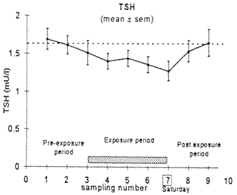
Figure 25: A significant reduction in Thyrotropin (Thyroid Stimulating Hormone) during cell phone use, de Seze et al. (1998).
Cell Site Health Surveys:
There is overwhelming evidence that cell sites are likely to cause a wide range of serious adverse health effects. Carefully designed health surveys are need to disprove or confirm this claim. Careful survey design includes consideration of exposure levels and patterns, as well as consideration of indoor and outdoor exposure levels that contribute differently to mean exposure levels.
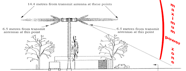
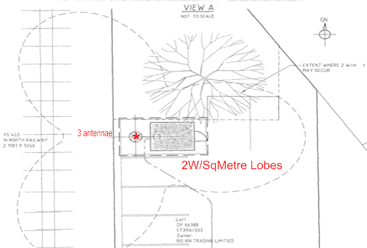
Cell site antennas focus most of the radiation into the main beam in the horizontal and vertical directions. The vertical antenna pattern includes two or three main side-lobes that produce the near tower ground level radiation exposures, Figures 26 to 28.
Figure 26: Cell site profile showing the extent of the
main beans and side lobes in which the 200 m W/cm2
standard is exceeded. This illustrates the directions of the beams and side lobes - (and the cone shaped area of greatest interest to
operators of nearby schools and playgrounds).
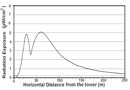
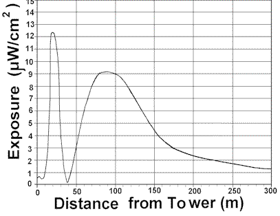
Cell site exposures for a low and high power sites are given in Figures 27 and 28. The side-lobes produce the nearer level and then the side of the main beam produces a wider peak and then falls off with distance from the tower. These two figures show the maximum exposure levels along the main beam direction. Figure 29 shows the horizontal pattern of a three-antenna tower radiation. The area between the main beam directions has a much lower exposure than in the main beam direction.
Figure 27: A low-powered cell site such as proposed for the Elmwood site.
Figure 28: A high-powered site as used at the Opawa Road site.
Figure 29: Three-panel horizontal radiation pattern, for a low powered site, as for the Elmwood Site.
Conclusions:
To over 40 studies have shown adverse biological or human health effects specifically from cell phone radiation. These research results to date clearly show that cell phones and cell phone radiation are a strong risk factor for all of the adverse health effects identified for EMR because they share the same biological mechanisms. The greatest risk is to cell phone users because of the high exposure to their heads and the great sensitivity of brain tissue and brain processes. DNA damage accelerates cell death in the brain, advancing neurodegenerative diseases and brain cancer. Brain tumour is already an identified risk factor. Cell phones are carried on people's belts and in breast pockets. Hence liver cancer, breast cancer and testicular cancer became probable risk factors.
Because the biological mechanisms for cell phone radiation mimics that of EMR, and the dose-response relationships have a threshold of ZERO, and this includes genetic damage, there is extremely strong evidence to conclude that cell sites are risk factors for:
A recommended risk reduction target for the mean chronic public exposure is 10 nW/cm2.
This is accomplished by setting the outside boundary exposure as 0.1m W/cm2.
References:
Abelin, T., 1999: "Sleep disruption and melatonin reduction from exposure to a shortwave radio signal". Seminar at Canterbury Regional Council, New Zealand. August 1999.
Adey, W.R., Byus, C.V., Cain, C.D., Higgins, R.J., Jones, R.A., Kean, C.J., Kuster, N., MacMurray, A., Stagg, R.B., Zimmerman, G., Phillips, J.L. and Haggren, W., 1999: "Spontaneous and nitrosourea-induced primary tumors of the central nervous system in Fischer 344 rats chronically exposed to 836 MHz modulated microwaves". Radiation Research 152(3): 293-302.
Alberts, E.N., 1977: "Light and electron microscopic observations on the blood-brain barrier after microwave irradiation. In Symposium on Biological effects and measurement of Radio Frequency/Microwaves, HEW Publication (FDA) 77-8026, pp 294-309.
Alberts, E.N., 1978: "Reversibility of microwave induced blood-brain barrier permeability". Radio Science Supplement.
Altpeter, E.S., Krebs, Th., Pfluger, D.H., von Kanel, J., Blattmann, R., et al., 1995: "Study of health effects of Shortwave Transmitter Station of Schwarzenburg, Berne, Switzerland". University of Berne, Institute for Social and Preventative Medicine, August 1995.
Altamura G, Toscano S, Gentilucci G, Ammirati F, Castro A, Pandozi C, Santini M, 1997: "Influence of digital and analogue cellular telephones on implanted pacemakers". Eur Heart J 18(10): 1632-4161.
Arnetz, B.B. and Berg, M., 1996: "Melatonin and Andrenocorticotropic Hormone levels in video display unit workers during work and leisure. J Occup Med 38(11): 1108-1110.
Balcer-Kubiczek, E.K. and Harrison, G.H., 1991: "Neoplastic transformation of C3H/10T1/2 cells following exposure to 120Hz modulated 2.45 GHz microwaves and phorbol ester tumor promoter". Radiation Research, 125: 65-72.
Balode, Z., 1996: "Assessment of radio-frequency electromagnetic radiation by the micronucleus test in Bovine peripheral erythrocytes". The Science of the Total Environment, 180: 81-86.
Barbaro V, Bartolini P, Donato A, Militello C, 1996: "Electromagnetic interference of analog cellular telephones with pacemakers". Pacing Clin Electrophysiol 19(10): 1410-1418.
Baris, D. and Armstrong, B., 1990: "Suicide among electric utility workers in England and Wales". Br J Indust Med 47:788-789.
Bawin, S.M. and Adey, W.R., 1976: "Sensitivity of calcium binding in cerebral tissue to weak electric fields oscillating at low frequency". Proc. Natl. Acad. Sci. USA, 73: 1999-2003.
Beall, C., Delzell, E., Cole, P., and Brill, I., 1996: "Brain tumors among electronics industry workers". Epidemiology, 7(2): 125-130.
Blackman, C.F., Benane, S.G., Elliott, D.J., and Pollock, M.M., 1988: "Influence of Electromagnetic Fields on the Efflux of Calcium Ions from Brain Tissue in Vitro: A Three-Model Analysis Consistent with the Frequency Response up to 510 Hz". Bioelectromagnetics, 9:215-227.
Blackman, C.F., Kinney, L.S., House, D.E., and Joines, W.T., 1989: "Multiple power-density windows and their possible origin". Bioelectromagnetics, 10: 115-128.
Blackman, C.F., 1990: "ELF effects on calcium homeostasis". In "Extremely low frequency electromagnetic fields: The question of cancer", BW Wilson, RG Stevens, LE Anderson Eds, Publ. Battelle Press Columbus: 1990; 187-208.
Borbely, AA, Huber, R, Graf, T, Fuchs, B, Gallmann, E, Achermann, P, 1999: Pulsed high-frequency electromagnetic field affects human sleep and sleep electroencephalogram. Neurosci Lett 275(3):207-210.
Bortkiewicz, A., Zmyslony, M., Palczynski, C., Gadzicka, E. and Szmigielski, S., 1995: "Dysregulation of autonomic control of cardiac function in workers at AM broadcasting stations (0.738-1.503 MHz)". Electro- and Magnetobiology 14(3): 177-191.
Bortkiewicz, A., Gadzicka, E. and Zmyslony, M., 1996: "Heart rate in workers exposed to medium-frequency electromagnetic fields". J Auto Nerv Sys 59: 91-97.
Bortkiewicz, A., Zmyslony, M., Gadzicka, E., Palczynski, C. and Szmigielski, S., 1997: "Ambulatory ECG monitoring in workers exposed to electromagnetic fields". J Med Eng and Tech 21(2):41-46.
Braune, S, Wrocklage, C, Raczek, J, Gailus, T, Lucking, CH, 1998: Resting blood pressure increase during exposure to a radio-frequency electromagnetic field. Lancet 351(9119):1857-1858.
Brueve, R., Feldmane, G., Heisele, O., Volrate, A. and Balodis, V., 1998: "Several immune system functions of the residents from territories exposed to pulse radio-frequency radiation". Presented to the Annual Conference of the ISEE and ISEA, Boston Massachusetts July 1998.
Burch, J.B., Reif, J.S., Pittrat, C.A., Keefe, T.J. and Yost, M.G., 1997: "Cellular telephone use and excretion of a urinary melatonin metabolite". In: Annual review of Research in Biological Effects of electric and magnetic fields from the generation, delivery and use of electricity, San Diego, CA, Nov. 9-13, P-52.
Burch, J.B., Reif, J.S., Yost, M.G., Keefe, T.J. and Pittrat, C.A., 1998: "Nocturnal excretion of urinary melatonin metabolite among utility workers". Scand J Work Environ Health 24(3): 183-189.
Burch, J.B., Reif, J.S., Yost, M.G., Keefe, T.J. and Pittrat, C.A., 1999a: "Reduced excretion of a melatonin metabolite among workers exposed to 60 Hz magnetic fields" Am J Epidemiology 150(1): 27-36.
Burch, J.B., Reif, J.S. and Yost, M.G., 1999b: "Geomagnetic disturbances are associated with reduced nocturnal excretion of melatonin metabolite in humans". Neurosci Lett 266(3):209-212.
Burch, J.B., Reif, J.S., Noonan, C.W. and Yost, M.G., 2000:
"Melatonin metabolite levels in workers exposed to 60-Hz magnetic fields: work in
substations and with 3-phase conductors". J of Occupational and Environmental
Medicine, 42(2): 136-142.
Byus, C.V., Kartun, K., Pieper, S. and Adey, W.R., 1988: "Increased ornithine decarboxylase activity in cultured cells exposed to low energy modulated microwave fields and phorbol ester tumor promoters". Cancer research, 48(15): 4222-4226.
Cantor, K.P., Stewart, P.A., Brinton, L.A., and Dosemeci, M., 1995: "Occupational exposures and female breast cancer mortality in the United States". Journal of Occupational Medicine, 37(3): 336-348.
Chen WH, Lau CP, Leung SK, Ho DS, Lee IS, 1996: "Interference of cellular phones with implanted permanent pacemakers". Clin Cardiol 19(11): 881-886.
Cossarizza, A., Angioni, S., Petraglia, F., Genazzani, A.R., Monti, D., Capri, M., Bersani, F., Cadossi, R. and Franceschi, C., 1993: "Exposure to low frequency pulsed electromagnetic fields increases interleukin-1 and interleukin-6 production by human peripheral blood mononuclear cells". Exp Cell Res 204(2):385-387.
Daniells, C, Duce, I, Thomas, D, Sewell, P, Tattersall, J, de Pomerai, D, 1998: "Transgenic nematodes as biomonitors of microwave-induced stress". Mutat Res 399: 55-64.
Dasdag, S, Ketani, MA, Akdag, Z, Ersay, AR, Sar,i I, Demirtas ,OC, Celik, MS, 1999: Whole-body microwave exposure emitted by cellular phones and testicular function of rats. Urol Res 27(3):219-223.
Davis, R.L. and Mostofl, 1993: "Cluster of testicular cancer in police officers exposed to hand-held radar". Am. J. Indust. Med. 24: 231-233.
Deroche, M., 1971: " Etude des perturbations biologiques chez les techniciens O.R.T.F. dans certains champs electromagnetiques de haute frequence". Arch Mal. Prof, 32: 679-683.
De Mattei, M., Caruso, A., Traina, G.C., Pezzetti, F., Baroni, T., and Sollazzo, V., 1999: "Correlation between pulsed electromagnetic fields exposure time and cell proliferation increase in human osteosarcoma cell lines and human normal osteoblast cells in vitro". Bioelectromagnetics 20: 177-182.
De Pomerai, D., Daniells, C., David, H., Duce, I., Mutwakil, M., Thomas, D., Sewell, P., Tattersall, J., Jones, D., and candido, P., 2000: "Non-thermal heat-shock response to microwaves". Nature May 25,
de Seze R, Fabbro-Peray P, Miro L, 1998: GSM radiocellular telephones do not disturb the secretion of antepituitary hormones in humans. Bioelectromagnetics 19(5):271-8.
Dmoch, A. and Moszczynski, P., 1998: "Levels of immunoglobulin and subpopulations of T lymphocytes and NK cells in men occupationally exposed to microwave radiation in frequencies of 6-12GHz". Med Pr 49(1):45-49.
Dolk, H., Shaddick, G., Walls, P., Grundy, C., Thakrar, B., Kleinschmidt, I. and Elliott, P., 1997a: "Cancer incidence near radio and television transmitters in Great Britain, I - Sutton-Colfield transmitter". American J. of Epidemiology, 145(1):1-9.
Dolk, H., Elliott, P., Shaddick, G., Walls, P., Grundy, C., and Thakrar, B.,1997b: "Cancer incidence near radio and television transmitters in Great Britain, II All high power transmitters". American J. of Epidemiology, 145(1):10-17.
Donnellan M, McKenzie DR, French PW, 1997: Effects of exposure to electromagnetic radiation at 835 MHz on growth, morphology and secretory characteristics of a mast cell analogue, RBL-2H3. Cell Biol Int 21:427-439.
Eulitz, C, Ullsperger, P, Freude, G, Elbert ,T, 1998: Mobile phones modulate response patterns of human brain activity. Neuroreport 9(14):3229-3232.
Fesenko, EE, Makar, VR, Novoselova, EG, Sadovnikov, VB, 1999: Microwaves and cellular immunity. I. Effect of whole body microwave irradiation on tumor necrosis factor production in mouse cells. Bioelectrochem Bioenerg 49(1):29-35.
Forman, S.A., Holmes, C.K., McManamon, T.V., and Wedding, W.R., 1982: "Physiological Symptoms and Intermittent Hypertension following acute microwave exposure". J. of Occup. Med. 24(11): 932-934.
Freude, G, Ullsperger, P, Eggert ,S, Ruppe, I, 1998: Effects of microwaves emitted by cellular phones on human slow brain potentials. Bioelectromagnetics 19(6):384-387.
French PW, Donnellan M, McKenzie DR, 1997: Electromagnetic radiation at 835 MHz changes the morphology and inhibits proliferation of a human astrocytoma cell line. Bioelectrochem Bioenerg 43:13-18.
Freude, G, Ullsperger, P, Eggert, S, Ruppe, I, 2000: Microwaves emitted by cellular telephones affect human slow brain potentials. Eur J Appl Physiol 81(1-2):18-27.
Frey, A.H., Feld, S.R. and Frey. B., 1975: "Neural function and behavior: defining the relationship in biological effects of nonionizing radiation". Ann. N.Y. Acad. Sci. 247: 433-438.
Frey, A.H., 1998: "Headaches from cellular telephones: are they real and what are the impacts". Environ Health Perspect 106(3):101-103.
Fritze K, Wiessner C, Kuster N, Sommer C, Gass P, Hermann DM, Kiessling M, Hossmann KA, 1997: Effect of global system for mobile communication microwave exposure on the genomic response of the rat brain. Neuroscience 81(3):627-639.
Garaj-Vrhovac, V., Fucic, A, and Horvat, D., 1990: "Comparison of chromosome aberration and micronucleus induction in human lymphocytes after occupational exposure to vinyl chloride monomer and microwave radiation"., Periodicum Biologorum, Vol 92, No.4, pp 411-416.
Garaj-Vrhovac, V., Horvat, D. and Koren, Z., 1991: "The relationship between colony-forming ability, chromosome aberrations and incidence of micronuclei in V79 Chinese Hamster cells exposed to microwave radiation". Mutat Res 263: 143-149.
Garaj-Vrhovac, V., Fucic, A, and Horvat, D., 1992: The correlation between the frequency of micronuclei and specific aberrations in human lymphocytes exposed to microwave radiation in vitro". Mutation Research, 281: 181-186.
Garaj-Vrhovac, V., and Fucic, A., 1993: "The rate of elimination of chromosomal aberrations after accidental exposure to microwave radiation". Bioelectrochemistry and Bioenergetics, 30:319-325.
Garaj-Vrhovac, V., 1999: "Micronucleus assay and lymphocyte mitotic activity in risk assessment of occupational exposure to microwave radiation. Chemosphere 39(13): 2301-2312.
Goldsmith, J.R., 1997a: "TV Broadcast Towers and Cancer: The end of innocence for Radiofrequency exposures". Am. J. Industrial Medicine 32 : 689-692.
Goldsmith, J.R., 1997b: "Epidemiologic evidence relevant to radar (microwave) effects". Environmental Health Perspectives, 105 (Suppl 6): 1579-1587.
Gordon, Z.V., 1966: "Problems of industrial hygiene and the biological effects of electromagnetic superhigh frequency fields". Moscow Medicina [In Russian] English translation in NASA Rept TT-F-633, 1976.
Goswami, P.C., Albee, L.D., Parsian, A.J., Baty, J.D., Moros, E.G., Pickard, W.F., Roti Roti, J.L. and Hunt, C.R., 1999: "Proto-oncogene mRNA levels and activities of multiple transcription factors in C3H 10T 1/2 murine embryonic fibroblasts exposed to 835.62 and 847.74 MHz cellular telephone communication frequency radiation". Radiat Res 151(3): 300-309.
Graham, C., Cook, M.R., Cohen, H.D. and Gerkovich, M.M., 1994: "A dose response study of human exposure to 60Hz electric and magnetic fields". Bioelectromagnetics 15: 447-463.
Graham, C., Cook, M.R., Sastre, A., Riffle, D.W. and Gerkovich, M.M., 2000: "Multi-night exposure to 60 Hz magnetic fields: effects on melatonin and its enzymatic metabolite". J Pineal Res 28(1): 1-8.
Grayson, J.K., 1996: "Radiation Exposure, Socioeconomic Status, and Brain Tumour Risk in the US Air Force: A nested Case-Control Study". American J. of Epidemiology, 143 (5), 480-486.
Haider, T., Knasmueller, S., Kundi, M, and Haider, M., 1994: "Clastogenic effects of radiofrequency radiation on chromosomes of Tradescantia". Mutation Research, 324:65-68.
Hamburger, S., Logue, J.N., and Sternthal, P.M., 1983: "Occupational exposure to non-ionizing radiation and an association with heart disease: an exploratory study". J Chronic Diseases, Vol 36, pp 791-802.
Hammett and Edison Inc., 1997: "Engineering analysis of radio frequency exposure conditions with addition of digital TV channels". Prepared for Sutra Tower Inc., San Francisco, California, January 3, 1997.
Hanson Mild, K, Oftedal, G, Sandstrom, M, Wilen, J, Tynes, T, Haugsdal, B, Hauger E, 1998: Comparison of symptoms experienced by users of analogue and digital mobile phones: a Swedish-Norwegian epidemiological study. Arbetslivsrapport 23.
Hardell, L, Reizenstein, J, Johansson, B, Gertzen, H, Mild, KH, 1999: Angiosarcoma of the scalp and use of a cordless (portable) telephone. Epidemiology 10(6):785-786.
Hardell, L, Nasman, A, Pahlson, A, Hallquist, A, Hansson Mild, K, 1999: Use of cellular telephones and the risk for brain tumours: A case-control study. Int J Oncol 15(1):113-116.
Hardell, L, Nasman, A, Hallquist, A, 2000: "Case-control study of radiology work, medical X-ray investigations and use of cellular telephones as risk factors". J of General Medicine. <www.medscape.com/Medscape/GeneralMedicine/journal/2000/v02.n03/>
Hayes, R.B., Morris Brown, L., Pottern, L.M., Gomez, M., Kardaun, J.W.P.F., Hoover, R.N., O'Connell, K.J., Sutsman, R.E. and Nasser, J., 1990: Occupational and Risk for Testicular Cancer: A Case Control Study. International Journal of Epidemiology, 19, No.4, pp 825-831.
Heller, J.H., and Teixeira-Pinto, A.A., 1959: "A new physical method of creating chromosome aberrations". Nature, Vol 183, No. 4665, March 28, 1959, pp 905-906.
Hladky, A, Musil, J, Roth, Z, Urban, P, Blazkova, V, 1999: Acute effects of using a mobile phone on CNS functions. Cent Eur J Public Health 7(4):165-167.
Hocking, B., Gordon, I.R., Grain, H.L., and Hatfield, G.E., 1996: "Cancer incidence and mortality and proximity to TV towers". Medical Journal of Australia, Vol 165, 2/16 December, pp 601-605.
Hocking, B, 1998: Preliminary report: symptoms associated with mobile phone use. Occup Med (Lond);48(6):357-360.
Hofgartner F, Muller T, Sigel H, 1996: "Could C- and D-network mobile phones endanger patients with pacemakers?". Dtsch Med Wochenschr 121(20): 646-652,. [Article in German]
Ivaschuk, O.I., Jones, R.A., Ishida-Jones, T., Haggren, Q., Adey, W.R. and Phillips, J.L., 1997: "Exposure of nerve growth factor-treated PC12 rat pheochromscytoma cells to a modulated radiofrequency field at 836.55 MHz: effects on c-jun and c-fos expression". Bioelectromagnetics 18(3): 223-229.
Jacobson, C.B., 1969: Progress report on SCC 31732, (Cytogenic analysis of blood from the staff at the U.S. Embassy in Moscow), George Washington University, Reproductive Genetics Unit, Dept. of Obstertics and Genocolgy, February 4, 1969.
Johnson-Liakouris, A.J.. 1998: "Radiofrequency (RF) Sickness in the Lilienfeld Study: an effect of modulated microwaves". Arch Environ Heath 53(3):236-238.
Jones, L.F., 1933: "A study of the propagation of wavelengths between three and eight meters. Proc. of the Institute of Radio Engineers 21(3): 349-386.
Jordan, E.C., (Ed), 1985: "Reference data for engineers: Radio, Electronics, Computer and Communications, 7th Edition". Publ. Howard W. Sams & CO., Indianapolis.
Juutilainen, J., Stevens, R.G., Anderson, L.E., Hansen, N.H., Kilpelainen, M., Laitinen, J.T., Sobel, E. and Wilson, B.W., 2000: "Nocturnal 6-hydroxymelatonin sulphate excretion in female workers exposed to magnetic fields". J Pineal Res 28(2): 97-104.
Kallen, B., Malmquist, G., and Moritz, U., 1982: "Delivery Outcome among Physiotherapists in Sweden: is Non-ionizing Radiation a Fetal Hazard? Archives of Environmental Health, 37(2): 81-84.
Karasek, M., Woldanska-Okonska, M., Czernicki, J., Zylinska, K. and Swietoslawski, J., 1998: "Chronic exposure to 2.9 mT, 40 Hz magnetic field reduces melatonin concentrations in humans". J Pineal Research 25(4): 240-244.
Kellenyi, L, Thuroczy, G, Faludy, B, Lenard, L, 1999: Effects of mobile GSM radiotelephone exposure on the auditory brainstem response (ABR). Neurobiology 7:79-81.
Khudnitskii, SS, Moshkarev, EA, Fomenko, TV, 1999: [On the evaluation of the influence of cellular phones on their users]. [Article in Russian] Med Tr Prom Ekol (9):20-24.
Kolomytkin, O., Kuznetsov, V., Yurinska, M, Zharikova, A., and Zharikov, S., 1994: "Response of brain receptor systems to microwave energy exposure". pp 195-206 in "On the nature of electromagnetic field interactions with biological systems", Ed Frey, A.H., Publ. R.G. Landes Co.
Koivisto, M, Revonsuo, A, Krause, C, Haarala, C, Sillanmaki, L, Laine, M, Hamalainen, H, 2000: Effects of 902 MHz electromagnetic field emitted by cellular telephones on response times in humans. Neuroreport 11(2):413-415.
Kolodynski, A.A. and Kolodynska, V.V., 1996: "Motor and psychological functions of school children living in the area of the Skrunda Radio Location Station in Latvia". The Science of the Total Environment, Vol 180, pp 87-93.
König HL. 1974, Behavioural changes in human subjects associated with ELF electric fields. In Persinger MA, editor. ELF and VLF electromagnetic field effects. New York, Plenum Press.
Krause, C.M., Sillanmaki, L., Koivisto, M., Haggqvist, A., Saarela, C., Revonsuo, A., Laine, M. and Hamalainen H., 2000: "Effects of electromagnetic field emitted by cellular phones on the EEG during a memory task". Neuroreport 11(4): 761-764.
Kwee, S, Raskmark, P, 1997: Radiofrequency electromagnetic fields and cell proliferation. Presented at the Second World Congress for Electricity and Magnetism in Biology and Medicine, Bologna, Italy, June.
Lai, H. and Singh, N.P., 1995: "Acute low-intensity microwave exposure increases DNA single-strand breaks in rat brain cells". Bioelectromagnetics, Vol 16, pp 207-210, 1995.
Lai, H. and Singh, N.P., 1996: "Single- and double-strand DNA breaks in rat brain cells after acute exposure to radiofrequency electromagnetic radiation". Int. J. Radiation Biology, 69 (4): 513-521.
Lai, H., and Singh, N.P., 1997: "Melatonin and Spin-Trap compound Block Radiofrequency Electromagnetic Radiation-induced DNA Strands Breaks in Rat Brain Cells." Bioelectromagnetics, 18:446-454.
Lamble D, Kauranen T, Laakso M, Summala H, 1999: "Cognitive load and detection thresholds in car following situations: safety implications for using mobile (cellular) telephones while driving". Accid Anal Pre ;31(6):617-623.
Larsen, A.I., Olsen, J., and Svane, O., 1991: "Gender specific reproductive outcome and exposure to high frequency electromagnetic radiation among physiotherapists". Scand. J. Work Environ. Health, Vol.17, pp 324-329.
Lilienfeld, A.M., Tonascia, J., and Tonascia S., Libauer, C.A., and Cauthen, G.M., 1978: "Foreign Service health status study - evaluation of health status of foreign service and other employees from selected eastern European posts". Final Report (Contract number 6025-619073) to the U.S. Dept of State, July 31, 1978.
Lilienfeld, A.M., 1983: "Practical limitations of epidemiologic method". Environmental Health Perspectives, 52:3-8.
Lindbohm, M-L,, Hietanen, M., Kyyronen, P., Sallmen, M., von Nandelstadh, P., Taskinen, H., Pekkarinen, M., Ylikoski, M. and Hemminki, K., 1992: "Magnetic fields of video display terminals and spontaneous abortion". Am J Epidemiol 136:1041-1051.
Litovitz, T.A., Krause, D., Penafiel, M., Elson, E.C. and Mullins, J.M., 1993: "The role of coherence time in the effect of microwaves on ornithine decarboxylase activity". Bioelectromagnetics 14(5): 395-403.
Maes, A., Verschaeve, L., Arroyo, A., De Wagter, C. and Vercruyssen, L., 1993: "In vitro effects of 2454 MHz waves on human peripheral blood lymphocytes". Bioelectromagnetics 14: 495-501.
Maes, A., Collier, M., Slaets, D., and Verschaeve, L., 1996: "954 MHz Microwaves enhance the mutagenic properties of Mitomycin C". Environmental and Molecular Mutagenesis, 28: 26-30.
Maes A, Collier M, Van Gorp U, Vandoninck S, Verschaeve L, 1997: Cytogenetic effects of 935.2-MHz (GSM) microwaves alone and in combination with mitomycin C. Mutat Res 393(1-2): 151-156.
Mann, K, Roschke, J, 1996: Effects of pulsed high-frequency electromagnetic fields on human sleep. Neuropsychobiology 33(1):41-47.
Maskarinec, G. Cooper, J. and Swygert, L., 1994: "Investigation of increased incidence in childhood leukemia near radio towers in Hawaii: Preliminary observations". J. Environ Pathol Toxicol and Oncol 13(1): 33-37.
McKenzie, D.R., Yin, Y. and Morrell, S., 1998: "Childhood incidence of acute lymhoblastic leukaemia and exposure to broadcast radiation in Sydney - a second look". Aust NZ J Pub Health 22 (3): 360-367.
Michelozzi, P., Ancona, C., Fusco, D., Forastiere, F. and Perucci, C.A., 1998: "Risk of leukamia and residence near a radio transmitter in Italy". ISEE/ISEA 1998 Conference, Boston Mass. Paper 354 P., Abstract in Epidemiology 9(4):S111.
Mild, K.H., Oftedal, G., Sandstrom, M., Wilen, J., Tynes, T., Haugsdal, B. and Hauger E., 1998: "Comparison of symptoms by users of analogue and digital mobile phones - A Swedish-Norwegian epidemiological study". National Institute for working life, 1998:23, Umea, Sweden, 84pp.
Milham, S., 1982: "Mortality from leukemia in workers exposed to electric and magnetic fields". New England J. of Med., 307: 249-250.
Milham, S., 1985: "Silent Keys", Lancet 1, 815, 1985.
Milham S., 1985: "Mortality in workers exposed to electromagnetic fields. Environ Health Perspectives 62:297-300.
Milham, S., 1988: "Increased mortality in amateur radio operators due to lymphatic and hematopoietic malignancies". Am. J. Epidemiology, Vol 127, No.1, pp 50-54.
Milham, S., 1996: "Increased incidence of cancer in a cohort of office workers exposed to strong magnetic fields". Am. J. Ind. Med. 30(6): 702-704.
Moscovici, B., Lavyel, A. and Ben Itzhac, D., 1974: "Exposure to electromagnetic radiation among workers". Family Physician 3(3): 121.
Moszczynski, P., Lisiewicz, J., Dmoch, A., Zabinski, Z., Bergier, L., Rucinska, M. and Sasiadek, U., 1999: "The effect of various occupational exposures to microwave radiation on the concentrations of immunoglobulins and T lymphocyte subsets". Wiad Lek 52(1-2):30-34.
Nakamura, H., Seto,T., Nagase, H., Yoshida, M., Dan, S. and Ogina, K., 1997: "Effects of exposure to microwaves on cellular immunity and placental steroids in pregnant rats. Occup Environ Med 54(9):676-680.
Naegeli B, Osswald S, Deola M, Burkart F, 1996: "Intermittent pacemaker dysfunction caused by digital mobile telephones". J Am Coll Cardiol 27(6):1471-1477.
Occhetta E, Plebani L, Bortnik M, Sacchetti G, Trevi G, 1999: "Implantable cardioverter defibrillators and cellular telephones: is there any interference?". Pacing Clin Electrophysiol 22(7): 983-989.
Oscar, K.J. and Hawkins, T.D., 1997: "Microwaves alteration of the blood-brain barrier system of rats". Brain Research 126: 281-293.
Ouellet-Hellstrom, R. and Stewart, W.F., 1993: "Miscarriages among Female Physical Therapists who report using radio- and microwave- frequency electromagnetic radiation." American J. of Epidemiology, 138 (10): 775-86.
Persson, B.R.R., Salford, L.G. and Brun, A., 1997: "Blood-brain barrier permeability in rats exposed to electromagnetic fields used in wireless communication". Wireless Network 3: 455-461.
Penafiel, L.M., Litovitz, T., Krause, D., Desta, A. and Mullins, J.M., 1997: "Role of modulation on the effect of microwaves on ornithine decarboxylase activity in L929 cells". Bioelectromagnetics 18(2): 132-141.
Perry, F.S., Reichmanis, M., Marino, A. and Becker, R.O., 1981: "Environmental power-frequency magnetic fields and suicide". Health Phys 41(2): 267-277.
Pfluger, D.M. and Minder, C.E., 1996: "Effects of 16.7 Hz magnetic fields on urinary 6-hydroxymelatonin sulfate excretion of Swiss railway workers". J Pineal Research 21(2): 91-100.
Phelan, A.M., Lange, D.G., Kues, H.A, and Lutty, G.A., 1992: "Modification of membrane fluidity in Melanin-containing cells by low-level microwave radiation". Bioelectromagnetics 13: 131-146.
Philips, J.L., Haggren, W., Thomas, W.J., Ishida-Jones, T. and Adey, W.R., 1992: "Magnetic field-induced changes in specific gene transcription". Biochem Biophys Acta 1132(2): 140-144.
Philips, J.L., Haggren, W., Thomas, W.J., Ishida-Jones, T. and Adey, W.R., 1993: "Effect of 72 Hz pulsed magnetic field exposure on ras p21 expression in CCRF-CEM cells". Cancer Biochem Biophys 13(3): 187-193.
Phillips, J.L., Ivaschuk, O., Ishida-Jones, T., Jones, R.A., Campbell-Beachler, M. and Haggnen, W.,1998: "DNA damage in molt-4 T-lymphoblastoid cells exposed to cellular telephone radiofrequency fields in vitro". Bioelectrochem Bioenerg 45: 103-110.
Pollack, H., 1979: "Epidemiologic data on American personnel in the Moscow Embassy", Bull. N.Y. Acad. Med, 55(11): 1182-1186.
Pollack, H., 1979a: "The microwave syndrome", Bull. N.Y. Acad. Med, 55(11): 1240-1243.
Preece, AW, Iwi, G, Davies-Smith, A, Wesnes, K, Butler, S, Lim, E, Varey, A, 1999: Effect of a 915-MHz simulated mobile phone signal on cognitive function in man. Int J Radiat Biol 75(4):447-456.
Quan, R., Yang, C., Rubinstein, S., Lewiston, N.J., Sunshine, P., Stevenson, D.K. and Kerner, J.A., 1992: "Effects of microwave radiation on anti-infective factors in human milk". Pediatrics 89(4):667-669.
Reiter, R.J., 1994: "Melatonin suppression by static and extremely low frequency electromagnetic fields: relationship to the reported increased incidence of cancer". Reviews on Environmental Health. 10(3-4): 171-86, 1994.
Reiter, R.J. and Robinson, J, 1995: "Melatonin: Your body's natural wonder drug". Publ. Bantam Books, New York.
Repacholi, MH, Basten, A, Gebski, V, Noonan, D, Finnie, J, Harris, AW, 1997: Lymphomas in E mu-Pim1 transgenic mice exposed to pulsed 900 MHZ electromagnetic fields. Radiat Res 147(5):631-640.
Robinette, C.D., Silverman, C. and Jablon, S., 1980: "Effects upon health of occupational exposure to microwave radiation (radar)". American Journal of Epidemiology, 112(1):39-53, 1980.
Rosen, L.A., Barber, I. and Lyle D.B., 1998: "A 0.5 G, 60 HZ magnetic field suppresses melatonin production in pinealocytes". Bioelectromagnetics 19: 123-127.
Rothman KJ, Loughlin JE, Funch DP, Dreyer NA.,1996: Overall mortality of cellular telephone customers. Epidemiology 7:303-305.
Sagripanti, J. and Swicord, M.L., 1976: DNA structural changes caused by microwave radiation. Int. J. of Rad. Bio., 50(1), pp 47-50, 1986.
Salford, L.G., Brun, A., Sturesson, K., Eberhardt, J.L. and Persson, B.R.R., 1994: Permeability of the Blood-Brain Barrier induced by 915 MHz electromagnetic radiation, continuous wave and modulated at 8, 16, 50 and 200 Hz.
Sarkar, S., Ali S, and Behari, J., 1994: "Effect of low power microwave on the mouse genome: A direct DNA analysis". Mutation Research, 320: 141-147.
Savitz, D.A., Checkoway, H. and Loomis, D.P., 1998a: "Magnetic field exposure and neurodegenerative disease mortality among electric utility workers". Epidemiology 9(4):398-404.
Savitz, D.A,, Loomis, D.P. and Tse, C.K., 1998b: "Electrical occupations and neurodegenerative disease: analysis of U.S. mortality data". Arch Environ Health 53(1): 71-74.
Savitz, D.A., Liao, D., Sastre, A., Klecjner, R.C., and Kavet, R., 1999: "Magnetic field exposure and cardiovascular disease mortality among electric utility workers". Am. J. Epidemiology, 149(2): 135-142.
Schlegel RE, Grant FH, Raman S, Reynolds D 1998: "Electromagnetic compatibility study of the in-vitro interaction of wireless phones with cardiac pacemakers". Biomed Instrum Technol 32(6): 645-655.
Schwartz,, J.L., House, D.E., and Mealing, A.R., 1990: "Exposure of frog hearts to CW or amplitude modulated VHF fields: selective efflux of calcium ions at 16 Hz." Bioelectromagnetics, 11: 349-358.
Selvin, S., Schulman, J. and Merrill, D.W., 1992: "Distance and risk measures for the analysis of spatial data: a study of childhood cancers". Soc. Sci. Med., 34(7):769-777.
Shandala, M.G., Dumanskii, U.D., Rudnev, M.I., Ershova, L.K., and Los I.P., 1979: "Study of Non-ionising Microwave Radiation Effects on the Central Nervous System and Behavior Reactions". Environmental Health Perspectives, 30:115-121.
Sobel, E., Davanipour, Z., Sulkava, R., Erkinjuntti, T., Wikstrom, J., Henderson, V.W., Bucjwalter, G., Bowman, D. and Lee, P-J., 1995: "Occupations with exposure to electromagnetic fields: a possible risk factor for Alzheimers Disease". Am J Epidemiol 142(5): 515-524.
Sobel, E., Dunn, M., Davanipour, D.V.M., Qian, M.S. and Chui, M.D., 1996: "Elevated risk of Alzheimers disease among workers with likely electromagnetic field exposure. Neurology 47(12): 1477-1481.
Schirmacher, A, Bahr, A, Kullnick, U, Stoegbauer, F, 1999: Electromagnetic fields (1.75 GHz) influence the permeability of the blood-brain barrier in cell culture model. Presented at the Twentieth Annual Meeting of the Bioelectromagnetics Society, St. Pete Beach, FL, June.
Stark, K.D.C., Krebs, T., Altpeter, E., Manz, B., Griol, C. and Abelin, T., 1997: "Absence of chronic effect of exposure to short-wave radio broadcast signal on salivary melatonin concentrations in dairy cattle". J Pineal Research 22: 171-176.
Szmigielski, S., 1996: "Cancer morbidity in subjects occupationally exposed to high frequency (radiofrequency and microwave) electromagnetic radiation". Science of the Total Environment, Vol 180, 1996, pp 9-17.
Szmigielski, S., Bortkiewicz, A., Gadzicka, E., Zmyslony, M. and Kubacki, R., 1998: "Alteration of diurnal rhythms of blood pressure and heart rate to workers exposed to radiofrequency electromagnetic fields". Blood Press. Monit, 3(6): 323-330.
Thomas, T.L., Stolley, P.D., Stemhagen, A., Fontham, E.T.H., Bleecker, M.L., Stewart, P.A., and Hoover, R.N., 1987: "Brain tumor mortality risk among men with electrical and electronic jobs: A case-control study". J. Nat. Canc. Inst., Vol 79, No.2, pp 233-238., August 1987.
Tice R, Hook G, McRee DI., 1999: "Chromosome aberrations from exposure to cell phone radiation". Microwave News, Jul/Aug (1999) p7.
Timchenko, O.I., and Ianchevskaia, N.V., 1995: "The cytogenetic action of electromagnetic fields in the short-wave range". Psychopharmacology Series, Jul-Aug;(7-8):37-9.
Trigano AJ, Azoulay A, Rochdi M, Campillo, A., 1999: "Electromagnetic interference of external pacemakers by walkie-talkies and digital cellular phones: experimental study. Pacing Clin Electrophysiol 22(4 Pt 1): 588-593.
Tonascia, J.A. and Tonascia, S., 1969: "Hematological Study: progress report on SCC 31732", George Washington University, Department of Obstectrics and Gynecology, February 4, 1969.
Tynes, T., Hannevik, M., Anderson, A., Vistnes, A.I. and Haldorsen, T., 1996: "Incidence of breast cancer in Norewegian female radio and telegraph operators". Cancer causes Control., 7(2): 197-204.
Van Wijngaarden, E., Savitz, D.A., Kleckner, R.C., Dai, J. and Loomis, D., 2000: "Exposure to electromagnetic fields and suicide among electric utility workers: a nested case-control study". Occupational and Environ Medicine, 57: 258-263.
Vaneeck, J., 1998: "Cellular Mechanisms of Melatonin Action". Physiol. Rev. 78: 687-721.
Velizarov, S, Raskmark, P, Kwee, S, 1999: The effects of radiofrequency fields on cell proliferation are non-thermal. Bioelectrochem Bioenerg 48(1):177-180.
Verschaeve, L., Slaets, D., Van Gorp, U., Maes, A. and Vanderkom, J., 1994: "In vitro and in vivo genetic effects of microwaves from mobile phone frequencies in human and rat peripheral blood lymphocytes". Proceedings of Cost 244 Meetings on Mobile Communication and Extremely Low Frequency field: Instrumentation and measurements in Bioelectromagnetics Research. Ed. D, Simunic, pp 74-83.
Vijayalaxmi, B.Z., Frei, M.R., Dusch, S.J., Guel, V., Meltz, M.L. and Jauchem, J.R., 1997: "Frequency of micronuclei in the peripheral blood and bone marrow of cancer-prone mice chronically exposed to 2450 MHz radiofrequency radiation". Radiation Research, 147: 495-500.
Violanti, J.M., 1998: "Cellular phones and fatal traffic collisions". Accid Anal Prev 30(4): 519-524.
Violanti, J.M. and Marshall, J.R., 1996: "Cellular phones and traffic accidents: an epidemiological approach". Accid Anal Prev 28(2): 265-270.
Von Klitzing, L, 1995: Low-frequency pulsed electromagnetic fields influence EG of man. Phys. Medica 11:77-80.
Youbicier-Simo, BJ, Lebecq, JC, Bastide, M, 1998: Mortality of chicken embryos exposed to EMFs from mobile phones. Presented at the Twentieth Annual Meeting of the Bioelectromagnetics Society, St. Pete Beach, FL, June.
Walleczek, J., 1992: "Electromagnetic field effects on cells of the immune system: the role of calcium signaling". FASEB J., 6: 3176-3185.
Wang, S.G. 1989: "5-HT contents change in peripheral blood of workers exposed to microwave and high frequency radiation". Chung Hua Yu Fang I Hsueh Tsa Chih 23(4): 207-210.
Wever R. 1974, ELF-effects on Human Circadian Rhythms. In: Persinger MA editor. ELF and VLF Electromagnetic Field Effects. New York, Plenum Press. p 101-144.
Weyandt, T.B., Schrader, S.M., Turner, T.W. and Simon, S.D., 1996: "Semen analysis of military personnel associated with military duty assignments". Reprod Toxicol 10(6):521-528.
Wilson, B.W., Wright, C.W., Morris, J.E., Buschbom, R.L., Brown, D.P., Miller, D.L., Sommers-Flannigan, R. and Anderson, L.E., 1990: "Evidence of an effect of ELF electromagnetic fields on human pineal gland function". J Pineal Research 9(4): 259-269.
Wood, A.W., Armstrong, S.M., Sait, M.L., Devine, L. and Martin, M.J., 1998: "Changes in human plasma melatonin profiles in response to 50 Hz magnetic field exposure". J Pineal Research 25(2): 116-127.
Zaret, M.M., 1977: "Potential hazards of hertzian
radiation and tumors. NY State J Med,146-147.
To return to orange index click HERE
.
This site is evolving therefore you may wish to use refresh button each visit.